









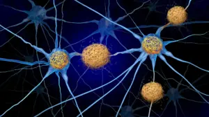

In a groundbreaking study that challenges decades of conventional wisdom regarding Alzheimer’s disease, researchers at the University of Kentucky have uncovered an unexpected reason why the condition so severely disrupts sleep. Their findings suggest that the persistent exhaustion and sleep disturbances experienced by patients may not be a direct result of the disease’s hallmark protein deposits, but rather a byproduct of the brain’s own defensive immune response. By targeting this immune reaction in laboratory mice, scientists were able to restore more than two hours of sleep per day without ever removing the amyloid plaques typically associated with the pathology.
The research, recently published in the journal Alzheimer’s & Dementia, was led by Shannon L. Macauley, Ph.D., an associate professor of physiology at the University of Kentucky College of Medicine, alongside first author Nicholas J. Constantino, Ph.D., a recent doctoral graduate. The discovery is being hailed as "paradigm-shifting," as it pivots the focus of sleep research away from damaged neurons or plaque accumulation and toward the complex, often counterproductive, behavior of the brain’s specialized immune cells, known as microglia.
How the Brain’s Immune System Can Disrupt Sleep
To understand the mechanics of this discovery, it is helpful to use the analogy of a domestic fire. Imagine a small flame breaking out in a kitchen. A perfectly calibrated fire suppression system would target the heat source directly. However, in the context of Alzheimer’s disease, it appears the brain’s "sprinkler system"—the microglia—activates with overwhelming intensity, flooding the entire house and causing far more collateral damage than the initial fire.

Amyloid plaques, which are sticky deposits of protein fragments, have long been the primary focus of Alzheimer’s research. They accumulate between nerve cells and are considered a hallmark of the disease. While microglia are intended to protect the nervous system by clearing debris and responding to injury, they can become overzealous when they detect these plaques. Instead of merely containing the protein deposits, these cells release a cascade of inflammatory signals. According to Dr. Macauley, these inflammatory molecules interfere with the brain’s normal activity, essentially keeping the "lights on" in the brain when it should be resting. "Microglia are immune cells that, when they respond to plaques, kick off this elaborate cascade of inflammation, as if the microglia are partying all night and keeping the brain awake," she explains.
This "whole house response" suggests that the immune system’s reaction to Alzheimer’s pathology is a major, yet previously overlooked, driver of clinical symptoms. By shifting the focus from the plaques themselves to the inflammatory environment they trigger, researchers have opened a new door for potential therapeutic interventions.
Tracking Alzheimer’s Sleep Changes Inside the Brain
The investigation involved a rigorous comparison between two groups of mice: those genetically predisposed to develop amyloid plaques and a control group of "wild-type" mice that experienced the typical, gradual effects of aging. By studying these animals at six months—when plaques first emerge—and at 18 months, when the disease pathology is advanced, the team could isolate the specific impacts of Alzheimer’s from the background noise of natural aging.
To capture high-fidelity data, the team utilized a combination of electroencephalography (EEG) and electromyography (EMG). The EEG recorded the rhythmic electrical oscillations of the brain, while the EMG monitored muscle activity. This dual-monitoring approach allowed researchers to precisely map the sleep stages of the mice, identifying exactly when and how their rest was being interrupted.
Furthermore, the team employed light-sheet microscopy, a sophisticated imaging technique that renders biological tissue transparent. By illuminating the brain with a thin laser sheet, they created detailed three-dimensional maps of the brain, allowing them to visualize the exact spatial distribution of both amyloid plaques and the reactive microglia. This provided crucial evidence of where the immune cells were clustering in response to the disease.
Scientists Temporarily Remove 87% of Brain Immune Cells
The pivotal moment in the study occurred when the researchers sought to confirm that microglia were the true drivers of the sleep disturbances. They administered a drug known as Pexidartinib (PLX3397), which is known to block the survival signals upon which microglia depend. After 14 days of treatment, the researchers successfully depleted approximately 87% of the microglia in the mice.
To ensure the resulting data was accurate, the team utilized a mathematical method called Fitting Oscillations and One Over Frequency (FOOOF). This technique allowed the scientists to separate the brain’s electrical signals into periodic waves—the rhythm of brain activity—and aperiodic, background "noise." This allowed them to determine if the brain was continuing to fire at high intensity even when the animal was theoretically at rest. The findings confirmed that when the microglia were absent, the "engine" of the brain finally slowed down, allowing for significantly better sleep.
A Surprising Discovery About Alzheimer’s Brain Plaques
One of the most mind-blowing results of the study was the revelation of a "ceiling effect" regarding sleep disruption. The team had initially hypothesized that as the plaque burden grew, the quality of sleep would decline in a linear, proportional fashion. Instead, they found that sleep problems emerged early in the disease process and remained relatively stable, even as the amount of amyloid in the brain more than doubled by the 18-month mark.
"I expected that as plaque burden became more severe, sleep disruption would also worsen," noted Dr. Constantino. The data suggested that the initial inflammatory "spark" caused by the first plaques is enough to trigger the sleep-disrupting inflammatory cascade, and further accumulation of plaque does not necessarily exacerbate the sleep loss further. This implies that the early-stage immune response is a critical window for potential treatment.
Alzheimer’s Disrupts a Different Type of Sleep Than Normal Aging
The study further clarified that the nature of sleep disruption in Alzheimer’s is distinct from the sleep changes seen in healthy aging. While normal aging typically results in a reduction of REM sleep—the stage associated with dreaming and memory consolidation—Alzheimer’s pathology specifically targets non-rapid eye movement (NREM) sleep.
NREM sleep is the deep, physically restorative phase where the brain performs essential maintenance, such as flushing out metabolic waste. Dr. Macauley emphasized the importance of this stage, noting, "That restorative sleep is super important for physical repair, learning and memory and washing out the toxins of the day." The loss of this stage creates a dangerous feedback loop: the pathology disturbs sleep, and the resulting lack of deep sleep prevents the brain from clearing the very proteins that may be fueling the disease in the first place.
More Than Two Hours of Sleep Restored Without Removing Plaques
The most striking outcome of the experiment was that removing the microglia resulted in an increase of more than two hours of sleep per day for the mice, specifically improving the duration of deep, restorative NREM sleep. Most importantly, this was achieved without the removal of a single amyloid plaque.
This finding fundamentally changes the narrative of Alzheimer’s treatment. It demonstrates that the disease’s most disruptive symptoms may be managed by targeting the immune system’s inflammatory response, rather than solely focusing on the challenging task of clearing amyloid deposits. While this is an experimental breakthrough in mice, it offers a hopeful roadmap for future human trials.
The Research Team Behind the Discovery
The success of this study was rooted in the collaborative culture fostered at the University of Kentucky’s Sanders-Brown Center on Aging. Dr. Macauley, who emphasizes a philosophy of "calculated risk-taking," credits the project’s success to her team’s willingness to challenge long-held assumptions. The environment in her laboratory encourages students to prioritize curiosity over dogma. "I love people who take initiative, find their passion, are curious, and keep pushing to find an answer," Macauley said.
Dr. Constantino, reflecting on his time in the lab, noted that the most significant breakthroughs often occurred when their original hypotheses proved incorrect. "Dr. Macauley has also taught me to embrace uncertainty and failure as part of the scientific process," he said. This focus on "following the data" is what ultimately led the team to look past the plaques and discover the role of the brain’s immune system.
Could Existing Medications Help Restore Sleep in Alzheimer’s?
Looking toward the future, the research team is exploring how to translate these mouse-model findings into human therapies. Because microglia are essential for basic brain health, eliminating them entirely is not a viable strategy for human patients. Instead, the team is investigating ways to "calm" the overactive cells.
They are currently testing whether existing, FDA-approved medications—such as the diabetes drug Metformin or the anti-seizure drug Stiripentol—might modulate microglial metabolism. By changing how these cells produce and consume energy, the researchers hope to reduce their inflammatory output without destroying the cells themselves. The goal is to intervene early in the disease process, potentially improving the cognitive health and daily quality of life for patients before memory loss becomes severe.
A Simpler Way to Detect Early Alzheimer’s Changes
Beyond treatment, the study highlighted the potential of EEG as a powerful diagnostic tool. Because the team identified specific electrical signatures associated with the inflammatory response to Alzheimer’s, they believe portable, affordable EEG systems could eventually be used to monitor patients in their own homes.
This could be a game-changer for rural populations, such as those in Kentucky, who may lack easy access to specialized medical centers and high-cost imaging. By using EEG as a "longitudinal biomarker," doctors could potentially screen for early signs of Alzheimer’s-related sleep disruption years before more invasive symptoms manifest. While these applications require further validation in human clinical trials, the research provides a clear, actionable path toward a more accessible and effective approach to understanding and treating the early stages of Alzheimer’s disease.