








Most primates are characterized by their remarkably complex brains, which are typically defined by a convoluted surface of deep grooves and ridges. This distinctive folding, known as gyrification, is a hallmark of intelligence and advanced cognitive function in species ranging from great apes to humans. However, the common marmoset (Callithrix jacchus) stands out as a curious evolutionary outlier. Despite being a primate, this small, South American monkey possesses a brain that is almost entirely smooth, lacking the intricate folds that scientists have long associated with higher-order processing power.
Now, researchers at the German Primate Center (DPZ), the Leibniz Institute for Primate Research in Göttingen, have pinpointed the precise cellular mechanisms that differentiate the marmoset brain from its more folded counterparts. By utilizing miniature, three-dimensional brain models known as organoids, the research team has uncovered a series of developmental shifts—involving the structure, division speed, and timing of neural progenitor cells—that effectively limit the production of neurons in marmosets, resulting in a smaller, smoother cortex. The findings, recently published in the journal Science Advances, offer a profound look into the evolutionary pathways of the primate brain and hold significant implications for our understanding of human developmental disorders.
Why Some Primate Brains Are Smooth Instead of Folded
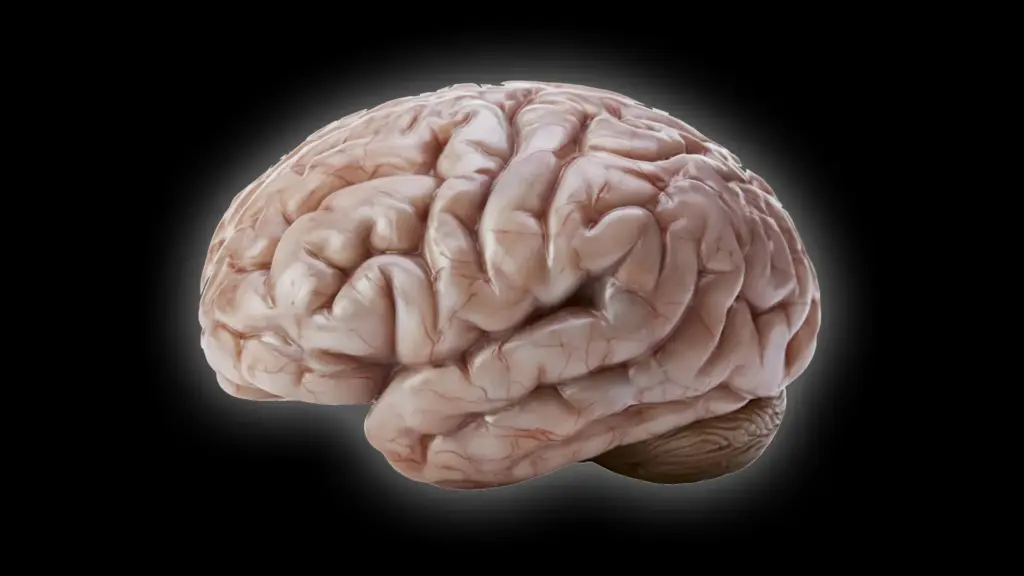
The evolutionary trajectory of the primate brain has been marked by a constant trade-off between size, efficiency, and surface architecture. The human brain, often described as resembling a walnut due to its high degree of folding, utilizes these convolutions to pack a vast amount of gray matter into the confined space of the cranium. This surface expansion is essential, as it provides the physical real estate required for billions of neurons to form the intricate networks that underpin complex problem-solving, emotional regulation, and advanced mental capabilities.

Historically, evolutionary biologists have operated under the assumption that the common ancestor of modern primates possessed a moderately sized brain that already exhibited some level of folding. Yet, as primates diverged into the diverse species we recognize today, this trait was not uniformly conserved. The common marmoset, an increasingly vital subject in modern biomedical research due to its manageable size and well-mapped genetics, has moved away from this pattern. Their brains are characterized by a near-total absence of visible folds, a mystery that has long intrigued neuroscientists.
To solve this puzzle, the researchers at the German Primate Center sought to identify the cellular "brakes" that prevent the marmoset brain from developing the same degree of complexity as that of a human. "We wanted to understand which changes at the cellular level cause the marmoset brain to grow less and form fewer folds," explains Lidiia Tynianskaia, a PhD student in the Junior Research Group Brain Development and Evolution at the DPZ and a co-first author of the study.
The investigation revealed a surprising starting point: at the very dawn of development, the common marmoset brain actually mimics the foundational structure of a large, folded primate brain. The divergence is not present from the start, but rather emerges as the brain matures. "As development progresses, processes must therefore occur that effectively slow down the production of nerve cells," Tynianskaia notes. This realization shifted the focus of the research from the early formation of the brain to the specific developmental windows where growth patterns begin to deviate.
Scientists Discover What Slows Brain Growth
To map these developmental differences, the research team employed brain organoids, which serve as laboratory-grown models that recapitulate key features of early brain development. By comparing organoids derived from human cells with those derived from common marmoset cells, the scientists could observe the activity of neural progenitor cells in real-time. These cells are the architects of the brain; they are responsible for generating the neurons that populate the cerebral cortex. The logic is simple but profound: the more neurons these progenitors can produce, the more the cortex can expand, and the greater the likelihood that the brain will fold to accommodate that growth.
The study revealed that the marmoset’s relatively smooth cerebral cortex is not the byproduct of a single, isolated genetic mutation. Instead, it is the result of a multi-faceted developmental program where several types of progenitor cells interact to limit the final number of neurons produced. "Our investigations have shown that certain progenitor cells in the common marmoset divide significantly more slowly than in humans," explains César Mateo Bastidas Betancourt, also a first author of the study and a PhD student in the Junior Research Group.
Beyond the rate of division, the study highlighted structural deficiencies. The researchers observed that marmoset progenitor cells possess a simpler architecture compared to their human counterparts, featuring fewer cellular processes. This structural simplicity correlates directly with lower proliferative capacity. When progenitor cells cannot divide rapidly or frequently, the pool of available neurons shrinks, leading to a smaller overall cortical volume that lacks the pressure or necessity to form folds.
Furthermore, the timing of these processes emerged as a critical factor. The research team discovered that the window of time during which these progenitor cells can multiply rapidly is significantly shorter in marmosets than in humans. This abbreviated period of growth acts as a final barrier to brain expansion, ensuring that the marmoset brain remains compact and smooth. Together, these factors—slower division, structural simplicity, and a shorter developmental timeline—create a powerful, compounded effect that restricts the growth of the marmoset brain.
Miniature Brains Reveal Important Developmental Clues
The use of organoids was fundamental to the success of this research. These miniature, three-dimensional tissues allowed the team to conduct experiments that would be statistically impossible or ethically prohibitive in living primates. By growing these models in a controlled environment, the scientists could track the precise developmental timing of key cellular processes across numerous samples.
"The study combines the advantages of in vivo and in vitro methods," says Michael Heide, head of the Junior Research Group Brain Development and Evolution. "Organoids are well-suited for obtaining statistically robust results because such sample sizes are not feasible in primates. We subsequently repeated some key experiments in fetal brain tissue to confirm the results from the organoids."
One of the most vital technical achievements of the study was bridging the gap between the laboratory model and the biological reality of the marmoset. Because organoids and actual brains do not always develop at the same chronological speed, the researchers had to establish a developmental benchmark. They discovered that a marmoset brain organoid grown for 50 days correlates precisely with the developmental stage of a natural marmoset brain at 90 days. Establishing this "chronological map" was essential; it allowed the scientists to ensure that they were comparing apples to apples, providing a reliable framework for interpreting how cellular behaviors in a dish translate to the biological outcomes observed in nature.
What Smooth Monkey Brains Reveal About Human Brain Evolution
The implications of this study extend far beyond the neurobiology of the common marmoset. By demonstrating how subtle, incremental changes in the behavior of progenitor cells can lead to drastic variations in brain anatomy, the researchers have provided a clearer picture of how primate brains have evolved over millions of years. This research suggests that the wide spectrum of brain shapes and sizes found among primates today is not necessarily the result of radical, high-level genetic overhauls, but rather the result of fine-tuning the cellular "clocks" and "building blocks" of early development.
For evolutionary biologists, this offers a compelling narrative on the diversification of the primate order. For medical researchers, the potential applications are equally significant. Understanding the precise cellular mechanisms that control neuron production provides a baseline for investigating human neurodevelopmental disorders. When these fundamental growth processes are disrupted in humans, the consequences can be severe, leading to conditions characterized by abnormal brain size or structural irregularities.
By identifying the specific cellular behaviors that "shrink" the marmoset brain, researchers have gained a new set of markers to look for when studying human conditions. The work conducted by the team at the German Primate Center underscores the value of comparative biology; by studying why some brains are smooth, we are gaining the tools to better understand why others—specifically our own—are so complex, and how to intervene when that complexity goes awry. As the field continues to refine these organoid models, the path forward promises a deeper understanding of the biological architecture that makes the primate brain one of the most remarkable products of evolution.