







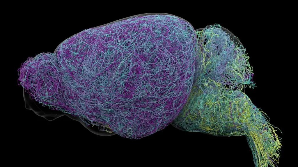
Deep within the brainstem, nestled in a region known as the locus coeruleus (LC)—Latin for "blue place"—lies a small cluster of nerve cells that exerts a disproportionate influence over our daily existence. Despite representing only a tiny fraction of the brain’s total neuronal population, this region serves as the primary engine for the production of norepinephrine (NE). This vital chemical messenger is the architect of our alertness, the regulator of our heart rate, and the primary conductor of our physiological responses to stress, excitement, and cognitive demand.
For decades, the prevailing consensus in neuroscience has characterized the locus coeruleus as a biological "loudspeaker." Scientists assumed that when the LC fired, it broadcast a uniform, non-specific signal of norepinephrine across the entire brain, effectively alerting every corner of the nervous system to a change in state. This "foghorn" model suggested that the brain was being bathed in a generalized chemical bath, prompting a global reaction to stimuli.
However, a groundbreaking study published in the journal Nature has fundamentally dismantled this long-standing assumption. By utilizing a sophisticated suite of modern techniques—including advanced brain imaging, high-resolution neural recordings, cutting-edge genetic tracing, and meticulous behavioral experiments in mice—researchers at the Allen Institute for Brain Science have revealed that the locus coeruleus is far from a uniform broadcaster. Instead, the LC communicates through a highly organized, complex network of distinct pathways. Rather than issuing a one-size-fits-all alarm, it delivers precise, targeted signals to specific destinations, with specialized groups of neurons responding to unique experiences and environmental demands.
A Hidden Brain Network That Sends Precise Messages
The research team began by meticulously mapping the connections of individual LC neurons to understand how they interface with the broader nervous system. What they discovered was a highly structured architecture that belies the previous understanding of the region as a blunt instrument of signaling. The study revealed that different subpopulations of cells are mapped to specific regions of the nervous system, creating a "postal network" that dictates exactly where norepinephrine is released.
This internal organization is defined by anatomical geography. Neurons located in the upper, or dorsal, portion of the locus coeruleus were found to maintain strong, dedicated connections to the cerebral cortex and the forebrain—areas of the brain responsible for high-level cognition and executive function. Conversely, cells situated in the lower, or ventral, portion of the LC were shown to primarily project their signals downward to the brainstem and the spinal cord. This spatial arrangement allows the LC to exert influence over distinct brain functions through physically separated pathways, providing a level of granular control that was previously invisible to researchers.
"The findings suggest the brain’s norepinephrine system is far more like a targeted postal network than a foghorn," said Karel Svoboda, director of Neural Dynamics at the Allen Institute and a co-author of the study. This shift in perspective transforms how neuroscientists view the brain’s regulatory mechanisms, moving from a model of broad-spectrum arousal to one of localized circuit modulation.
Why the Discovery Could Matter for Brain Disorders
The implications of this discovery extend well beyond basic neurobiology; they offer a potential roadmap for the next generation of psychiatric and neurological medicine. Norepinephrine is already a well-established player in the regulation of attention, mood, and stress, and as such, it serves as a cornerstone target for a wide array of medications currently used to treat depression, anxiety, and Attention Deficit Hyperactivity Disorder (ADHD).
Furthermore, the locus coeruleus is among the very first regions to experience degradation in the progression of Alzheimer’s disease. The fact that these cells are so early to falter suggests that the dysfunction of these specific, targeted pathways may play a critical role in the cognitive decline associated with dementia.
The clinical hurdle has always been the "systemic" nature of current pharmaceutical interventions. Many drugs that modulate norepinephrine levels act on the entire brain, often leading to a therapeutic benefit in one domain while triggering unwanted side effects elsewhere. By establishing a clear, detailed map of the LC’s specialized pathways, researchers are now better positioned to develop future therapies that act with surgical precision. By targeting only the specific circuits involved in a patient’s particular symptoms—such as the cortical pathways for cognitive focus or brainstem pathways for autonomic regulation—scientists hope to minimize the "off-target" effects that often plague current psychiatric treatments. This research was made possible in part by support from the National Institutes of Health’s Brain Research Through Advancing Innovative Neurotechnologies (BRAIN) Initiative.
Scientists Identify Brain Cells Involved in Learning
As the researchers delved deeper into the functional differences between these neuronal groups, they uncovered a clear link between specific LC pathways and distinct behavioral states. The dorsal LC neurons, which connect to the cortex, proved to be particularly active during the process of learning. In experiments where mice were required to perform complex decision-making tasks, these dorsal neurons exhibited a significant increase in activity specifically when the animals were adjusting their strategies after encountering unfavorable results.
In contrast, the ventral LC neurons exhibited a completely different pattern of activity. Their signaling levels rose shortly before the mice chose to ignore environmental cues that signaled a potential reward. This suggests that the ventral portion of the LC plays a gatekeeping role, helping to regulate whether an animal engages with its environment or chooses to disengage.
"What emerged was a clear map: neurons in the dorsal LC that send signals upward to the cortex are involved in learning, whereas neurons in the ventral region projecting downward to the brainstem and spinal cord govern whether animals engage with their environment at all," Svoboda explained. Crucially, the researchers noted that these functional differences were matched by distinct gene expression patterns, confirming that the divide between these two populations is not just structural, but fundamental to their biological identity.
Researchers Discover a Record-Breaking Neuron
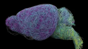
The scale of the study was immense, involving a comprehensive examination of the mouse brain that required massive data processing. The team utilized whole-brain imaging to examine nearly 35,000 neurons and analyzed the genetic profiles of almost 400,000 individual cells. Perhaps most strikingly, the researchers successfully reconstructed the complete, three-dimensional structures of several exceptionally complex nerve cells, revealing the physical extent of their influence.
During this reconstruction, the team encountered a surprising discovery regarding the reach of these cells. LC neurons possess extraordinarily long axons—the slender, cable-like projections that transmit electrical impulses to distant parts of the nervous system. The researchers found that, on average, these axons measured approximately 35 centimeters in length. One specific neuron stood out, with a total reconstructed length of 70.32 centimeters, marking it as one of the longest single neurons ever measured in a mouse.
"This neuron, like many others that we studied, supplies norepinephrine to a very large volume of the cerebral cortex," said Jeremiah Cohen, a scientist at the Allen Institute and study co-author. "For the brain, this is highly unusual. Most neurons are more specific in their targets." However, even this massive, far-reaching neuron was found to be highly selective. Despite its ability to span vast distances across the brain, it does not release norepinephrine universally; it pointedly ignores regions like the cerebellum, brainstem, and spinal cord. This reinforces the central thesis of the study: even the most widespread neurons in the LC are part of a carefully curated and organized system of signaling, rather than a disorganized distribution network.
A New Understanding of How the Brain Learns
The study also opened a new window into how the norepinephrine system interacts with other major chemical messengers in the brain, most notably dopamine. Dopamine is widely known for its role in the basal ganglia, a region essential for the formation of habits and the processing of rewards. Scientists now suspect that norepinephrine and dopamine may work in concert, forming a collaborative signaling framework that allows the brain to acquire and process multiple abstract concepts and complex behaviors simultaneously.
By mapping the intricate connections, specialized functions, and the surprising physical architecture of the locus coeruleus, this research provides a far more nuanced picture of how the brain manages the constant influx of information required for learning and survival. While these findings are derived from rodent models, the discovery of such a sophisticated, targeted signaling architecture offers a new lens through which to view human brain function. As researchers continue to refine this map, the path toward creating therapies that can selectively repair or adjust individual neural circuits—rather than simply saturating the brain with chemical signals—becomes increasingly clear. This study represents a significant leap forward in understanding the precise, elegant mechanisms that allow the brain to process the world around us.