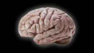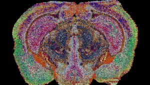
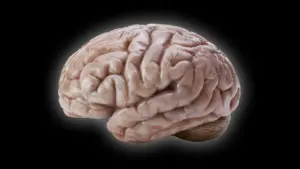








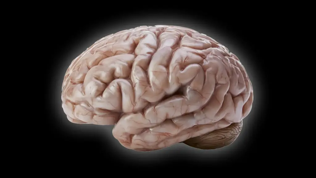
Most primates are characterized by their expansive, complex brains, which are typically defined by a surface topography of intricate folds and grooves. This architectural complexity is often cited as the biological foundation for the sophisticated cognitive abilities observed across the primate order. However, the common marmoset (Callithrix jacchus) stands out as a striking evolutionary anomaly. Despite being a primate, this small, South American monkey possesses a brain that is almost entirely lissencephalic—meaning it lacks the extensive folding seen in humans and other large-brained primates.
For years, the question of why some primates developed highly gyrified (folded) brains while others retained smooth surfaces has remained a subject of intense scientific inquiry. Now, a team of researchers at the German Primate Center (DPZ), the Leibniz Institute for Primate Research in Göttingen, has identified the specific cellular processes that govern these divergent developmental paths. By peering into the microscopic world of neural development, the researchers have illuminated how subtle differences in cell behavior can lead to profound differences in brain structure and size.
Why Some Primate Brains Are Smooth Instead of Folded
The evolutionary history of the primate brain is a tale of adaptation and diversification. The human brain, with its deeply folded surface resembling the ridges of a walnut, represents one end of a spectrum that allows for an enormous surface area within the confined space of the skull. This expansion provides the necessary real estate for billions of neurons, supporting the advanced mental faculties that define human intelligence. Evolutionary biologists generally posit that the common ancestor of modern primates possessed a moderately sized brain that likely featured at least some degree of folding. Yet, as primates evolved and diverged into different ecological niches, these characteristics shifted significantly.

The common marmoset has become an increasingly vital model organism in biomedical research due to its unique genetic and physiological traits. However, its brain structure is remarkably different from the human blueprint. To understand these differences, the research team at the DPZ sought to identify the cellular "instruction manual" that dictates brain growth.
"We wanted to understand which changes at the cellular level cause the marmoset brain to grow less and form fewer folds," explains Lidiia Tynianskaia, one of the study’s two first authors and a PhD student in the Junior Research Group Brain Development and Evolution at the DPZ. "At the beginning of development, the common marmoset brain exhibits the typical structure and composition of a large, folded primate brain. As development progresses, processes must therefore occur that effectively slow down the production of nerve cells."
Scientists Discover What Slows Brain Growth
To investigate these developmental nuances, the team employed a sophisticated methodology involving brain organoids—miniature, three-dimensional structures grown in the laboratory that mimic the early stages of brain development. By comparing organoids derived from human cells with those derived from marmoset cells, the scientists were able to observe the developmental trajectory of neural progenitor cells in real-time.
Neural progenitor cells are the architects of the brain; they are responsible for generating the neurons that populate the cerebral cortex. The size and shape of the mature brain are directly tied to the efficiency and output of these progenitor cells. A higher volume of neurons produced during fetal development provides the foundation for the cortex to expand and, eventually, to fold.
The findings, recently published in the journal Science Advances, indicate that the marmoset’s smoother cortex is not the result of a single genetic "switch." Instead, it is the product of a coordinated set of differences across several types of progenitor cells that work in tandem to limit neuronal output.
"Our investigations have shown that certain progenitor cells in the common marmoset divide significantly more slowly than in humans," explains César Mateo Bastidas Betancourt, the study’s co-first author and a PhD student in the Junior Research Group. "Other progenitor cells have a simpler structure than their human counterparts, with fewer processes, and are therefore less proliferative. Both of these factors ultimately result in fewer nerve cells, which contributes to a smaller size and less folding of the cerebral cortex in marmosets."
Furthermore, the researchers identified a critical temporal disparity. The window of time during which these progenitor cells can multiply rapidly is significantly shorter in marmosets than in humans. This abbreviated developmental period, combined with the slower rate of cell division and reduced structural complexity, creates a "bottleneck" that limits the total number of neurons produced.
Miniature Brains Reveal Important Developmental Clues
The use of brain organoids was pivotal to the success of the study. These models allowed the researchers to bypass the significant ethical and logistical hurdles associated with studying fetal primate brain development directly. By growing these tissues in a controlled environment, the team could perform statistically robust experiments that would be impossible to conduct in a traditional laboratory setting.
To ensure the validity of their findings, the researchers took the extra step of confirming their organoid-based observations against fetal brain tissue. "The study combines the advantages of in vivo and in vitro methods," says Michael Heide, head of the Junior Research Group Brain Development and Evolution. "Organoids are well-suited for obtaining statistically robust results because such sample sizes are not feasible in primates. We subsequently repeated some key experiments in fetal brain tissue to confirm the results from the organoids."
A significant challenge in organoid research is the discrepancy in growth rates between lab-grown models and natural biological development. To address this, the team meticulously calibrated their data, discovering that a marmoset brain organoid grown for 50 days acts as an accurate proxy for the developmental stage of a natural marmoset brain at 90 days. Establishing this chronological alignment was essential, as it allowed the researchers to make direct, reliable comparisons between the miniature models and the actual biological process, ensuring that the developmental milestones they observed were accurate reflections of reality.
What Smooth Monkey Brains Reveal About Human Brain Evolution
The implications of this research extend far beyond the anatomical curiosity of a smooth-brained monkey. By pinpointing the specific cellular mechanisms that constrain brain growth in the marmoset, the study provides a clearer picture of the evolutionary pressures that have shaped the diversity of primate brains. It demonstrates that the dramatic differences in brain morphology between species do not necessarily require radical genetic innovations; rather, they can emerge from subtle, incremental changes in the timing and rhythm of cellular development.
This insight into the "tuning" of brain development is of profound interest to neuroscientists studying human health. The processes that govern the proliferation of progenitor cells and the subsequent folding of the cortex are highly sensitive; when these processes go awry, they can lead to severe developmental disorders in humans. By understanding the healthy, albeit different, mechanisms that regulate brain size and structure in primates, researchers are better equipped to identify what goes wrong in cases of microcephaly, lissencephaly, and other neurodevelopmental conditions.
As the scientific community continues to explore the complex intersection of genetics and development, the common marmoset has proven to be an invaluable window into our own biology. What was once viewed as a simple anatomical difference between species has now been revealed as a sophisticated, multi-layered regulatory system. By decoding these processes, the DPZ researchers have not only advanced our understanding of how the primate brain evolved but have also laid a foundation for future breakthroughs in clinical neuroscience, potentially guiding the way toward new therapeutic interventions for brain development disorders in humans.