



For decades, the frontiers of neuroscience have been dominated by the study of the brain’s electrical signals, the architecture of its neuronal connections, and the complex expression of its genes and proteins. While these elements are undeniably central to our understanding of cognition and behavior, they represent only a portion of the biological story. Tucked away in the background—yet essential to every process from sensory perception to memory storage—lies a vast, largely uncharted landscape of lipids. These fatty molecules, which form the structural membranes surrounding neurons and provide the myelin insulation vital for signal transmission, have historically proven difficult to map. Now, a team of researchers at EPFL has bridged this significant knowledge gap, revealing a sophisticated chemical organization that challenges our existing perceptions of the brain.
The struggle to understand the brain’s lipid composition has long been a technical hurdle. Conventional imaging techniques, while effective for observing anatomy or gene expression, lack the resolution required to distinguish individual lipid molecules within the dense, crowded environment of neural tissue. This limitation left scientists with a blurry, generalized picture of the brain’s chemical makeup, masking the subtle variations that exist from one region to the next.
"Scientists have had only a blurry picture of how lipids differ from one part of the brain to another," explains Giovanni D’Angelo, professor at The Kristian Gerhard Jebsen Foundation Chair in Nutrition and Metabolism at EPFL. "That gap matters, because changes in brain lipids are increasingly linked to conditions from depression to Alzheimer’s disease."
The potential clinical implications of this research are profound. As the medical community seeks to understand the molecular roots of neurodegenerative and psychiatric disorders, the ability to pinpoint chemical irregularities in the brain could prove transformative. By establishing a high-resolution map of lipid distribution, the EPFL team, led by D’Angelo alongside colleagues Luca Fusar Bassini and Gioele La Manno, has provided the scientific community with a foundational tool to investigate how these fatty molecules shift in health and disease.
Identifying 539 Distinct Chemical Zones in the Brain
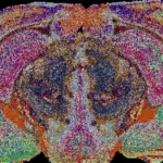
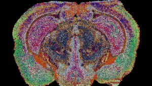
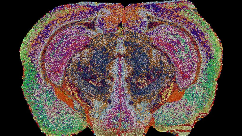
To construct what is now known as the Lipid Brain Atlas, the researchers employed mass spectrometry imaging—a sophisticated technique capable of identifying lipid species across ultra-thin sections of brain tissue. The process involved a laser scanning each slice of tissue at specific coordinates, meticulously cataloging the lipid profile of every location. By analyzing 172 different types of lipids across 109 brain slices collected from 11 mice, the team generated a massive dataset comprising approximately 7 million measurements. These data points were then synthesized into a comprehensive 3D reconstruction using specialized machine learning tools designed specifically for this project.
The result of this rigorous analytical process is a map revealing 539 chemically distinct territories, which the researchers have termed "lipizones." Each lipizone is defined by a unique combination of lipids, functioning as a recognizable chemical signature for that specific region of the brain.
The findings, recently published in the journal Nature, demonstrate that the brain’s chemical architecture is far more structured than previously assumed. Many of these lipizones align perfectly with established anatomical structures or known cell types, validating the accuracy of the technique. However, the atlas also uncovered unexpected relationships between groups of nerve cell bodies and distant, physically separated regions linked by their fibers. These functional connections, which have often been elusive when mapping via gene activity alone, become clear through the lens of lipid distribution.
"In effect, lipids work like postal codes, giving distant but related parts of the brain the same chemical address," D’Angelo notes. This "postal code" theory suggests that the brain uses lipid chemistry as a secondary, perhaps more subtle, method of organization to coordinate disparate regions that must function in harmony.
Hidden Chemical Patterns Challenge Previous Brain Maps
Perhaps the most surprising revelations from the Lipid Brain Atlas involve areas that scientists had previously characterized as uniform. A primary example is white matter, the dense tissue containing the nerve fibers that serve as the brain’s communication highway. While white matter is often depicted as a relatively homogenous connective tissue, the new atlas reveals that it is, in fact, composed of numerous distinct lipid zones.
"This shows that the cells which make myelin are far more varied in their chemistry than their genes alone would suggest," says Luca Fusar Bassini. This discovery implies that the oligodendrocytes—the cells responsible for producing myelin—might be operating under more nuanced regulatory controls than previously understood, potentially responding to localized chemical environments that are currently invisible to genetic mapping.
The team also identified previously unrecognized chemical boundaries within the choroid plexus, the specialized structure responsible for the production of cerebrospinal fluid, as well as in the tissues lining the brain’s internal cavities. These findings underscore the fact that lipid mapping provides a unique layer of biological data. It acts as a complementary resource, revealing features of brain organization that anatomy and genetics alone cannot illuminate, effectively allowing researchers to see the brain through a new, chemically defined prism.
Pregnancy Triggers Widespread Changes in Brain Lipids
Beyond merely mapping the static structure of the healthy brain, the atlas has provided a dynamic look at how the brain adapts to significant physiological events. By examining the lipid profiles of pregnant mice, the researchers discovered that pregnancy induces substantial, widespread shifts in lipid composition across numerous brain regions.
These changes were found to be more pronounced than the typical lipid variations observed between male and female mice, highlighting the intensity of the neurological adaptation required during pregnancy. The cortex—the outer layer of the brain responsible for high-level functions—showed the most extensive modifications. Furthermore, the team observed a sharp increase in galactosyl ceramide, a critical component of myelin, throughout the white matter.
These shifts suggest that the brain undergoes a systematic "retuning" of its internal wiring to meet the heightened metabolic and processing demands of pregnancy. As Gioele La Manno suggests, "The Atlas reveals that the brain constantly adjusts its lipids to meet the body’s changing needs." This suggests a level of neurochemical plasticity that scientists are only beginning to grasp, potentially explaining how the brain reconfigures itself to manage the complex physiological transitions associated with reproductive health.
A New Tool for Understanding Brain Aging and Disease
The Lipid Brain Atlas is now freely available online, offering the global scientific community a resource to explore the chemical nuances of the mouse brain. By integrating this lipid data with existing genetic and cellular atlases, researchers can now pursue a multidimensional understanding of brain organization, moving toward a more holistic view of how chemistry, structure, and gene expression interact over the lifespan.
The atlas serves as a definitive reference for the healthy mouse brain, providing a baseline against which pathological changes can be measured. For researchers investigating the onset of neurodegenerative diseases such as Alzheimer’s, or the chemical basis of mood disorders like depression, this tool offers a new pathway to identify how lipid distributions go awry. By monitoring how these "postal codes" shift during aging or in the presence of disease, scientists may be able to identify earlier biomarkers for conditions that currently lack effective early-detection methods.
While the current map is specific to the mouse brain, the researchers emphasize that the methodology is highly transferable. The success of this project proves that a highly organized lipid landscape is a fundamental feature of mammalian brain biology. The task ahead for the scientific community is to apply these techniques to human tissue samples.
"What transfers to humans isn’t the map, it’s the method, and the fact that we now know that a healthy brain has a very organized lipid map at all," says Fusar Bassini. With the groundwork laid, the field of neuro-lipidomics is poised to move from the margins of brain research to the forefront, promising a deeper, more chemically informed understanding of the organ that defines us.