









A good night’s sleep is often described as restorative, but the biological reality is far more dynamic than simply "recharging." While we slumber, the body engages in a complex, high-stakes physiological process that is essential for long-term health. During deep, non-REM sleep, the body releases growth hormone—a powerful chemical messenger that does far more than help teenagers reach their full height. It is a critical architect of the human body, responsible for building muscle, strengthening bones, and orchestrating the delicate process of fat metabolism.
For years, scientists have understood that sleep deprivation acts as a roadblock to this vital hormone’s production. However, the exact neural architecture—the "wiring" within the brain that triggers and controls this process—has remained a persistent mystery. Now, researchers at the University of California, Berkeley, have mapped the brain circuitry behind this relationship, offering a profound new understanding of how our sleep stages dictate our physical development and metabolic health.
In a landmark study published in the journal Cell, the UC Berkeley team identified the specific neurons that regulate growth hormone release. Perhaps more significantly, they discovered a previously unknown feedback loop that maintains a balance between hormone levels and our state of alertness. This discovery suggests that the relationship between sleep and growth hormone is a two-way street: sleep stimulates the production of the hormone, but the hormone, in turn, sends signals back to the brain that help dictate when we wake up.

The Hypothalamic Control Center
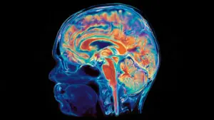
To unravel how the brain coordinates these disparate processes, the research team, led by Professor of Neuroscience and Molecular and Cell Biology Yang Dan, focused their investigation on the hypothalamus. Tucked deep within the brain, this small but mighty region serves as the body’s master control center. It is an evolutionary ancient structure, responsible for regulating hunger, body temperature, and the daily circadian rhythms of sleeping and waking.
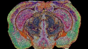
Within the hypothalamus, the researchers zoomed in on the neurons responsible for producing growth hormone-releasing hormone (GHRH) and two distinct types of neurons that produce somatostatin. These two chemical messengers act as a biological thermostat for growth hormone. GHRH serves as the accelerator, encouraging the pituitary gland at the base of the brain to secrete growth hormone into the bloodstream, while somatostatin acts as the brake, suppressing its release.
By analyzing how these neurons interact, the researchers began to see how the brain finely tunes the delivery of this hormone throughout the night. The team utilized electrodes implanted in mice to record neural activity, using light-based stimulation to observe how different parts of the hypothalamic system responded to specific triggers. Mice proved to be an ideal model for this research because their sleep architecture is fragmented—they sleep in short bursts throughout the day and night, providing researchers with repeated, real-time windows into the transition between wakefulness and sleep.
Distinct Patterns Across Sleep Stages
The study revealed that the interplay between GHRH and somatostatin is not static; it shifts dramatically depending on the stage of sleep. During REM (rapid eye movement) sleep, often associated with vivid dreaming, the brain increases activity in both GHRH and somatostatin neurons. While these hormones generally serve opposing roles, the researchers found that their coordinated activity during REM sleep actually contributes to an uptick in growth hormone secretion.
The pattern shifts again during non-REM sleep—the deep, restorative stage that typically dominates the earlier part of the night. In this state, somatostatin activity drops, effectively removing the "brake" on the system, while GHRH activity rises moderately. This shift creates the perfect chemical environment for sustained growth hormone release. By mapping these distinct neural patterns, the team has provided a clear explanation of how the brain manages to secrete the same essential hormone using entirely different biological mechanisms depending on the sleep stage.
The Growth Hormone Feedback Loop
Perhaps the most surprising finding in the study is that growth hormone does not simply sit at the receiving end of brain signals. It is an active participant in the sleep-wake cycle. As growth hormone levels accumulate during sleep, the hormone sends signals back to the brain, specifically targeting the locus coeruleus.
The locus coeruleus is a region in the brainstem that serves as a command post for alertness, attention, and the drive to explore new or unfamiliar environments. The researchers discovered that as growth hormone builds up, it stimulates neurons in the locus coeruleus, effectively promoting the transition from sleep to waking. This creates a sophisticated, self-regulating feedback loop: sleep drives the release of growth hormone, and that hormone then helps trigger the biological signal to wake up.
However, the system is fragile. The researchers identified a potential complication: while moderate stimulation of the locus coeruleus promotes healthy alertness, overstimulation can have a paradoxical effect. In an earlier study conducted by study co-author and UC Berkeley postdoctoral fellow Daniel Silverman, it was demonstrated that when these neurons become overexcited, they can actually trigger sleepiness rather than wakefulness. This suggests that the brain must maintain a delicate, homeostatic balance—a stable internal environment—to ensure that sleep remains restorative and that growth hormone levels do not disrupt our ability to stay awake.
Future Implications for Medicine
The clinical potential of these findings is significant. Because growth hormone plays a fundamental role in regulating glucose and fat metabolism, chronic sleep disruption—which interferes with this hormone’s release—is increasingly linked to a higher risk of obesity, diabetes, and cardiovascular disease. Furthermore, the locus coeruleus is known to be involved in various neurological and psychiatric conditions, including Parkinson’s and Alzheimer’s diseases.
"Understanding the neural circuit for growth hormone release could eventually point toward new hormonal therapies to improve sleep quality or restore normal growth hormone balance," Silverman explained. He noted that existing experimental gene therapies that target specific cell types could potentially be adapted to "dial back" the excitability of the locus coeruleus, offering a novel way to treat sleep-related disorders.
"People know that growth hormone release is tightly related to sleep, but that has historically been limited to drawing blood and checking levels," said lead author Xinlu Ding, a postdoctoral fellow in UC Berkeley’s Department of Neuroscience. "We’re actually directly recording neural activity in mice to see what’s going on. We are providing a basic circuit to work on in the future to develop different treatments."
The researchers emphasize that while the findings are promising, they are currently limited to models in mice. Further research is required to determine how these mechanisms function in human brains and whether such targeted therapies can be safely and effectively implemented.
Nonetheless, the study provides a foundational shift in how we view the hours we spend in bed. Rest is not a passive "shutting down" of the brain; it is a highly active, orchestrated period of physiological maintenance. The brain and body are in constant communication, using complex hormonal feedback loops to repair tissues, regulate metabolism, and prepare the mind for the demands of the coming day. By identifying the specific cells and signaling pathways that govern this process, the Berkeley team has opened a new door into the study of sleep, suggesting that the path to better health may lie in the very circuits that govern our deepest, most restorative moments of rest.
The research was supported by the Howard Hughes Medical Institute and the Pivotal Life Sciences Chancellor’s Chair fund. The study featured contributions from a broad team of researchers from UC Berkeley, including Peng Zhong, Bing Li, Chenyan Ma, Lihui Lu, Grace Jiang, Zhe Zhang, Xiaolin Huang, Xun Tu, and Zhiyu Melissa Tian, alongside Fuu-Jiun Hwang and Jun Ding of Stanford University.