








For decades, the scientific community has been obsessed with the hardware and the software of the human brain. Researchers have spent countless hours mapping the complex thickets of neurons, tracing the intricate connections of the connectome, and sequencing the genetic instructions that dictate brain development. Yet, as we have focused on these primary components, a vital piece of the biological puzzle has remained obscured in the shadows: the lipids. These diverse fatty molecules, which form the structural membranes surrounding neurons and provide the essential myelin insulation for our nerve fibers, have long been acknowledged as important, but until now, they have remained notoriously difficult to track.
A groundbreaking study led by researchers at EPFL (École Polytechnique Fédérale de Lausanne) has finally pulled back the curtain on this chemical landscape. By creating the first comprehensive, high-resolution map of lipid distribution in the mouse brain, scientists have discovered that the brain’s fatty architecture is not merely a uniform background, but a highly organized and distinct system. This new "Lipid Brain Atlas" promises to reshape our understanding of neurobiology, offering a new lens through which to view brain function, development, and the chemical shifts associated with conditions ranging from depression to Alzheimer’s disease.
The Hidden Chemical Geography of the Brain
For many years, the primary hurdle in lipid research was technological. While scientists understood that lipids were fundamental to brain function—acting as the building blocks for cell membranes and the signaling hubs for communication between neurons—conventional imaging techniques lacked the sensitivity to distinguish individual lipid molecules with any degree of spatial precision. The result was a blurry, generalized picture of brain chemistry that failed to capture how these molecules varied from one region to another.
"So scientists have had only a blurry picture of how lipids differ from one part of the brain to another," explains Giovanni D’Angelo, a professor at The Kristian Gerhard Jebsen Foundation Chair in Nutrition and Metabolism at EPFL. "That gap matters, because changes in brain lipids are increasingly linked to conditions from depression to Alzheimer’s disease."
To bridge this gap, a team led by D’Angelo, along with Luca Fusar Bassini and Gioele La Manno, embarked on a mission to map these molecules with unprecedented detail. The researchers utilized mass spectrometry imaging, a sophisticated technique that allows for the precise identification of molecules across microscopic sections of brain tissue. By employing a laser to scan each slice one location at a time, the team was able to pinpoint exactly which lipids were present and where they resided.
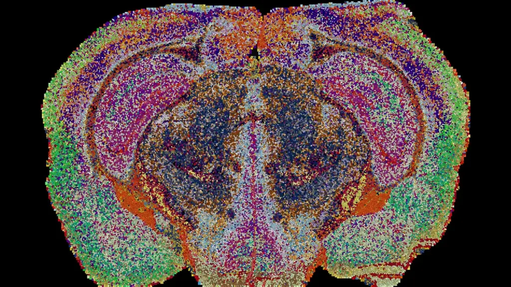
The scope of the project was immense. The team examined 172 distinct types of lipids across 109 brain slices harvested from 11 mice. This process generated approximately 7 million individual measurements, which were subsequently synthesized into a comprehensive 3D reconstruction using specialized machine learning tools developed specifically for this endeavor. The result of this massive computational effort was the Lipid Brain Atlas, a resource that revealed that lipids are not randomly dispersed. Instead, they are arranged in highly structured, recognizable patterns that often match known anatomical landmarks, while simultaneously revealing previously invisible chemical boundaries.
Defining the "Lipizones"
The most striking finding from the study, recently published in the journal Nature, is the identification of 539 chemically distinct territories within the mouse brain, which the researchers have dubbed "lipizones." Each of these territories possesses a unique combination of lipids, creating a specific chemical signature that is so distinctive that researchers can identify the origin of a brain tissue sample simply by analyzing its lipid composition.
Many of these lipizones align perfectly with established brain structures and specific cell types, confirming that the brain’s chemical organization is intimately tied to its anatomical architecture. However, the atlas also uncovered surprising relationships between groups of nerve cell bodies and distant, seemingly unrelated regions connected by their fibers. These connections have often proven difficult to detect using traditional maps based solely on gene activity, suggesting that lipids provide a form of "chemical signaling" that coordinates distant brain regions.
"In effect, lipids work like postal codes, giving distant but related parts of the brain the same chemical address," says D’Angelo. This discovery suggests that the brain’s organizational complexity extends far beyond what we can see through gene expression or cellular structure alone.
Challenging Conventional Maps
The Lipid Brain Atlas has also challenged long-held assumptions about the uniformity of brain tissue. For instance, white matter—the tissue composed of nerve fibers that act as the communication superhighways between brain regions—was previously thought to be relatively consistent in its composition. The new atlas reveals this to be a misconception; white matter is actually home to numerous, distinct lipid zones.
"This shows that the cells which make myelin are far more varied in their chemistry than their genes alone would suggest," notes Luca Fusar Bassini. This revelation suggests that the glia, or the supporting cells of the brain, are performing a much more nuanced role in regulating the environment of nerve fibers than previously understood. Furthermore, the researchers identified previously unrecognized chemical boundaries within the choroid plexus—the structure responsible for the production of cerebrospinal fluid—and in the tissues that line the brain’s internal cavities. These findings underscore the idea that lipid mapping can reveal critical features of brain organization that remain hidden when relying exclusively on traditional anatomical or genetic information.
Pregnancy and the Dynamic Brain
Perhaps the most compelling application of the new atlas is its ability to track how the brain changes in response to physiological states. When the researchers applied their mapping techniques to the brains of pregnant mice, they observed significant, widespread shifts in lipid composition. These changes were more pronounced than the typical lipid variations observed between male and female mice, highlighting the dynamic nature of the brain during this period.
The cortex, the outer layer of the brain associated with complex cognitive functions, showed particularly extensive lipid reorganization. Furthermore, levels of galactosyl ceramide, a crucial component of myelin, increased dramatically throughout much of the white matter. These observations suggest that pregnancy triggers a profound, systemic alteration in the brain’s chemical makeup.
"Together, these shifts suggest the brain retunes its own wiring to meet the demands of pregnancy," says Gioele La Manno. "The Atlas reveals that the brain constantly adjusts its lipids to meet the body’s changing needs." This indicates that the brain is not a static organ, but one that is constantly undergoing chemical recalibration to adapt to the body’s changing internal environment.
A New Era for Neurobiology
The implications of this work extend well beyond the laboratory mice used in the study. By making the Lipid Brain Atlas freely available online, the EPFL team has provided a transformative tool for the global scientific community. Researchers can now overlay lipid data onto existing genetic and cellular maps, creating a multi-dimensional view of the brain that is far more accurate than any single-discipline approach.
While the current map is specific to mice, the methodology behind it provides a roadmap for future human studies. By establishing a baseline for the healthy lipid distribution of a brain, scientists now have a reference point to investigate how these patterns are disrupted during development, aging, and the onset of neurological disorders. For example, if a specific lipid signature is associated with healthy cognitive function, researchers can more easily identify where those signatures break down in diseases like Alzheimer’s or depression.
"What transfers to humans isn’t the map, it’s the method, and the fact that we now know that a healthy brain has a very organized lipid map at all," says Fusar Bassini. By shifting the focus toward the "hidden" chemistry of the brain, this research opens a new frontier in neuroscience—one where the humble lipid is finally recognized as a central player in the complex drama of brain health and function. As the scientific community begins to explore this atlas, we are likely to find that the answers to some of the most persistent mysteries of the brain have been hiding in its fatty membranes all along.