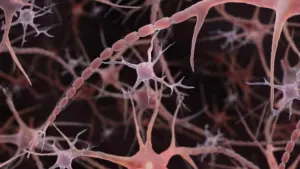









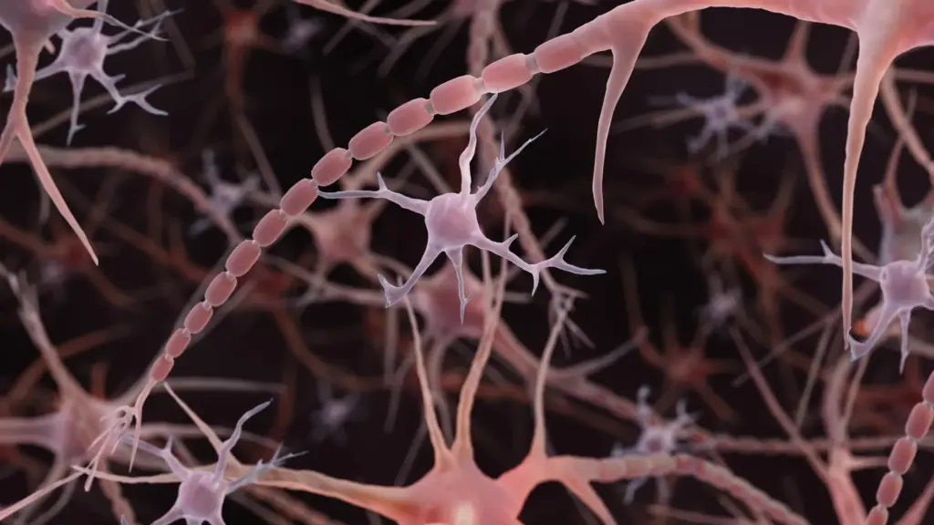
For over a century, the standard textbook illustration of a neuron has remained largely unchanged: a central cell body from which sprouts a long, slender, and uniformly smooth cable known as an axon. These axons, which act as the biological conduits for electrical impulses, have long been depicted as sleek, hollow tubes. However, a groundbreaking discovery by researchers at Johns Hopkins Medicine suggests that this iconic image is, at best, a simplification. Rather than being smooth, many axons actually resemble strings of tiny pearls, with repeating, nanoscale bulges that are far from merely decorative.
This unexpected architectural feature, unveiled in a study published online in Nature Neuroscience on December 2, 2024, indicates that the physical topography of the brain’s wiring is significantly more complex than previously assumed. By working with mouse neurons and employing advanced preservation techniques, the research team discovered that these "non-synaptic varicosities"—the technical term for these microscopic pearls—are a standard, functional component of healthy brain tissue.
A New Anatomy for Brain Cell Signaling
Axons are the fundamental infrastructure of the central nervous system, responsible for transmitting electrical signals from one neuron to another. Historically, the only "bulges" recognized in these structures were synaptic varicosities—the specialized sites where neurotransmitters are stored and released to bridge the gap between neurons. Any other form of beading observed in axons was traditionally dismissed as a sign of pathology. Indeed, in the context of neurodegenerative diseases such as Parkinson’s, pronounced axonal swelling is often a hallmark of cellular damage, reflecting a breakdown in the cell’s internal scaffolding and membrane integrity.

However, the Johns Hopkins study, led by Shigeki Watanabe, Ph.D., an associate professor of cell biology and neuroscience, shifted the paradigm by identifying much smaller, repeating swellings that occur in perfectly healthy neurons. Unlike the pathological beading associated with cell death, these "non-synaptic varicosities" appear to be an inherent part of the axon’s normal, healthy architecture.
"Understanding the structure of axons is important for understanding brain cell signaling," Watanabe noted in the university’s 2024 announcement. "Axons are the cables that connect our brain tissue, enabling learning, memory, and other functions. If our understanding of that cable’s shape is fundamentally incomplete, we must reconsider how we model the very signals that make us who we are."
The Role of Membrane Physics
The discovery of these beads traces back to Watanabe’s initial observations of repeating structures in the axons of worms. Seeking to understand the mechanics behind these shapes, Watanabe consulted with Swiss scientist Graham Knott, Ph.D. The researchers initially hypothesized that the axon’s internal skeleton—a complex network of proteins—might be responsible for the "pearled" appearance. However, when Jacqueline Griswold, a graduate student at Johns Hopkins and the study’s lead author, experimentally disrupted this skeletal framework, the pearls remained intact.
This result pushed the team to look elsewhere, leading them to collaborate with theoretical biophysicist Padmini Rangamani, Ph.D., a professor of pharmacology at the University of California San Diego School of Medicine. Together, the researchers hypothesized that the axon’s surrounding membrane, rather than its internal skeleton, was the primary architect of its shape.
To test this, the team employed high-pressure freezing electron microscopy. Standard imaging techniques often involve chemical fixation and dehydration, which can warp delicate cellular structures, turning a living, supple axon into a distorted, shriveled version of itself—much like the difference between a fresh grape and a raisin. By flash-freezing the tissue, the researchers were able to capture the neurons in a state closer to their living reality. Across tens of thousands of images of mouse neurons, the consistent, repeating pattern of pearling emerged, even in high-resolution images of living cells, confirming that the structure was not an artifact of the preparation process.
Mathematical Models and Membrane Tension
To explain why these pearls form, the team developed mathematical models of the axonal membrane. These simulations revealed that simple mechanical forces, such as membrane tension and stiffness, are sufficient to create these repeating swellings. The team’s experiments provided clear evidence for this: when they increased the concentration of sugar in the solution surrounding the axons, the swellings shrank. Similarly, increasing the membrane tension in the model resulted in smaller, more compressed pearls.
Cholesterol also emerged as a critical variable. When the researchers removed cholesterol from the membrane, the structure became less stiff and more fluid, which significantly altered the pearled appearance. More importantly, this change in structure directly impacted the speed of electrical conduction. "A wider space in the axons allows ions to pass through more quickly and avoid traffic jams," Watanabe explained. The findings suggest that the physical dimensions of these pearls and their connecting segments serve as a variable-speed regulator for neural communication.
Neural Activity and Structural Plasticity
Perhaps the most compelling finding is that these structures are not static. The team observed that when neurons were subjected to high-frequency electrical stimulation, the pearl-like regions underwent rapid and sustained changes. The bulges grew longer by an average of 8% and wider by 17%, with these structural modifications persisting for at least 30 minutes.
This activity-dependent change had a measurable effect on signaling, with electrical pulses slowing down significantly after stimulation. This effect lasted for up to an hour. Interestingly, when cholesterol was removed, these structural changes were blunted, and the characteristic slowing of the signal did not occur. This suggests that the axon is not merely a fixed, passive cable, but a dynamic, responsive structure capable of remodeling itself to tune the flow of information in real-time.
Beyond Mice: Observations in Human Tissue
Since the initial announcement of these findings in 2024, the scientific community has sought to determine if this "pearled" architecture is a universal feature of complex brains. A significant step forward occurred with a related study published in Neuron on November 24, 2025. Led by Chelsy R. Eddings and including Watanabe, this research documented the presence of pearled axons in human cortical tissue obtained during epilepsy surgeries.
By combining electrical stimulation with rapid freezing, the researchers successfully captured membrane dynamics in human brain slices that mirrored the findings in mice. While this does not mean every human axon displays identical pearling or that the functional consequences are uniform throughout the entire brain, it provides robust evidence that the "pearled" model is a biological reality rather than a species-specific anomaly. The study also highlighted the link between these structures and ultrafast endocytosis, the rapid recycling of cell membranes at synapses, further cementing the importance of membrane fluidity and shape in neural function.
A New Frontier in Neurobiology
The implications of this discovery are currently being explored through a major project funded by the National Institute of Mental Health (NIMH), which supports the continued collaboration between Watanabe and Rangamani. The project focuses on creating sophisticated computational models to predict how physical properties and external stimuli interact to shape axonal architecture.
Simultaneously, other research groups are developing new tools to investigate these microscopic dimensions. In July 2026, researchers at the University of Edinburgh published a study in PLOS Biology using automated imaging of living zebrafish to screen hundreds of compounds for their effects on axonal diameter. While this was not a direct replication of the Johns Hopkins pearling study, it identified 33 compounds that could influence the width of axons, providing a new toolkit for researchers to manipulate and study these structural features.
For the Johns Hopkins team, the ultimate goal remains the distinction between healthy, functional pearling and the damaging beading associated with neurodegenerative diseases. By clearly defining the parameters of "normal" axonal structure, researchers hope to better identify when and why the system begins to fail. As the field moves forward, the "smooth tube" model of the neuron is increasingly being replaced by a more nuanced, dynamic, and intricate view of the brain’s wiring—a view where the shape of the cable is just as important as the signal it carries.