



Deep brain stimulation (DBS) has long stood as one of the most transformative medical interventions for patients suffering from the debilitating motor symptoms of Parkinson’s disease. While the procedure—which involves the surgical implantation of electrodes to deliver precise electrical pulses to targeted deep-brain structures—is an established clinical standard, the precise neurological mechanisms that dictate its success have remained somewhat elusive. Now, an interdisciplinary team of researchers has provided a breakthrough, offering a clearer, more comprehensive picture of why this treatment works by identifying the specific brain network and the unique "rhythm" that appears to facilitate its therapeutic benefits.
The study, published in the prestigious journal Brain under the title "The Deep Brain Stimulation Response Network in Parkinson’s Disease Operates in the High Beta Band," represents a significant leap forward in the field of computational neurology. By synthesizing two methodologies that have historically been utilized in isolation—electrophysiology and advanced brain imaging—scientists from the University Hospitals of Cologne and Düsseldorf, Harvard Medical School, and Charité Berlin have effectively bridged the gap between spatial anatomy and temporal signaling.
Pinpointing Where and How Brain Stimulation Works
For decades, the medical community has grappled with the inherent limitations of studying the brain in pieces. Brain imaging studies have historically excelled at identifying the "where"—pinpointing the exact anatomical coordinates where electrical stimulation provides the most significant relief for tremors, rigidity, and bradykinesia. Conversely, electrophysiological research has focused on the "how"—the frequencies of the electrical signals that pulse through the brain’s circuitry. Until now, however, these two distinct lenses had rarely been unified to capture the full, synchronized picture of brain activity.
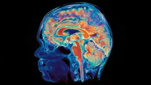
"For the first time, we were able to characterize the DBS response network in Parkinson’s disease in terms of space and time, simultaneously," explains Professor Dr. Andreas Horn, a specialist in computational neurology at the University of Cologne who led the research. "We show that Parkinson’s disease can best be treated if we stimulate a very precisely defined network. This network operates synchronized within a specific frequency band, and offers an explanation for how well patients respond to deep brain stimulation."
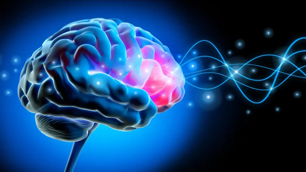
The core of the discovery lies in the recognition that the subthalamic nucleus—the primary target for DBS in Parkinson’s treatment—does not operate in a vacuum. Rather, it functions as a critical node within a complex, interconnected web of neural pathways. By mapping these connections, the researchers identified that the most effective therapeutic outcomes are inextricably linked to a network that communicates primarily through a relatively fast beta rhythm, specifically oscillating between 20 and 35 Hz.
Mapping a Parkinson’s Brain Network
To reach these conclusions, the international research consortium conducted a robust investigation involving a multicenter group of fifty patients and one hundred brain hemispheres. The scale of the study allowed for a high degree of statistical confidence, enabling the team to draw meaningful correlations between neuroanatomical data and clinical outcomes.
The methodology utilized in the study was particularly innovative. The researchers performed simultaneous recordings of brain activity using both the implanted DBS electrodes and magnetoencephalography (MEG). While the electrodes provided internal data from the depths of the brain, the MEG provided a non-invasive, high-resolution view of activity across the surface of the brain. By integrating these data streams, the team was able to map functional connections between the deep-brain regions and the frontal areas of the cerebral cortex.
The results of this mapping were definitive: the network connecting the subthalamic nucleus with frontal areas of the brain is not merely a passive conduit. It is an active communication channel that oscillates within the high beta band. Crucially, the researchers discovered that the strength of this specific connection was a strong predictor of patient outcomes. The patients whose stimulation protocols most effectively engaged this rhythmic network showed the most significant improvement in their motor symptoms following the electrode implantation. This suggests that the clinical success of DBS is not just about stimulating a general area, but about "tuning in" to a specific frequency that the brain uses for internal communication.
A Brain Rhythm That May Shape Treatment Response
The identification of this beta-band rhythm as a mediator of therapeutic effect opens new doors for the future of personalized medicine. As current DBS protocols rely on standardized settings, many patients may find that their relief is incomplete or that they require significant trial-and-error adjustments after the initial surgery.
"These results suggest that a certain rhythm of the brain acts as a communication channel between the subthalamic nucleus and the cerebral cortex and may mediate the therapeutic effects of deep brain stimulation," explains Dr. Bahne Bahners, the study’s first author, who is based at Düsseldorf University Hospital. "By stimulating regions that are connected to the identified network, we will probably be able to adjust DBS settings more precisely in the future, especially in patients who have not yet benefited optimally from deep brain stimulation."
This finding is particularly encouraging for the segment of the Parkinson’s population that experiences sub-optimal results with traditional DBS programming. If clinicians can visualize the patient’s individual network and understand how it responds to different stimulation parameters, they can move away from a "one-size-fits-all" approach to a more tailored, precision-medicine model. By targeting the nodes of this network that synchronize at the 20-35 Hz range, practitioners may be able to maximize symptom relief while potentially minimizing the power requirements of the device, thereby preserving battery life and reducing the likelihood of stimulation-related side effects.
The implications for clinical practice are profound. By providing a clear, measurable target—the high beta band rhythm—the research team has given clinicians a new metric to evaluate the efficacy of their programming. Rather than relying solely on the observation of motor symptoms, which can be subjective and time-consuming to assess, doctors may eventually be able to use the brain’s own electrical rhythm as a feedback mechanism to calibrate stimulation levels in real-time.
As the scientific community digests these findings, the research team is already looking toward the next phase of the project. While the current study establishes a powerful correlation between network connectivity, rhythmic signaling, and clinical improvement, the researchers are now moving to investigate the causality of these effects. Ongoing studies are currently focused on determining how exactly deep brain stimulation causes changes within these brain networks on a moment-to-moment basis. These causal studies are essential for understanding whether the stimulation simply "masks" the symptoms or if it induces a lasting, plastic change in the way the brain’s networks are organized.
The study, which was supported largely by funding from the Professor Klaus Thiemann Foundation, marks a turning point in the understanding of Parkinson’s disease management. By revealing the "language" of the network that DBS influences, the research does more than just explain why a treatment works—it paves the way for a new generation of neuro-technologies that are more precise, more effective, and more deeply attuned to the unique biological rhythms of the individual patient. As this field continues to evolve, the ability to read and modulate the brain’s internal oscillations may well become the gold standard for treating movement disorders, shifting the focus from general electrical stimulation to the fine-tuned orchestration of neural communication.