








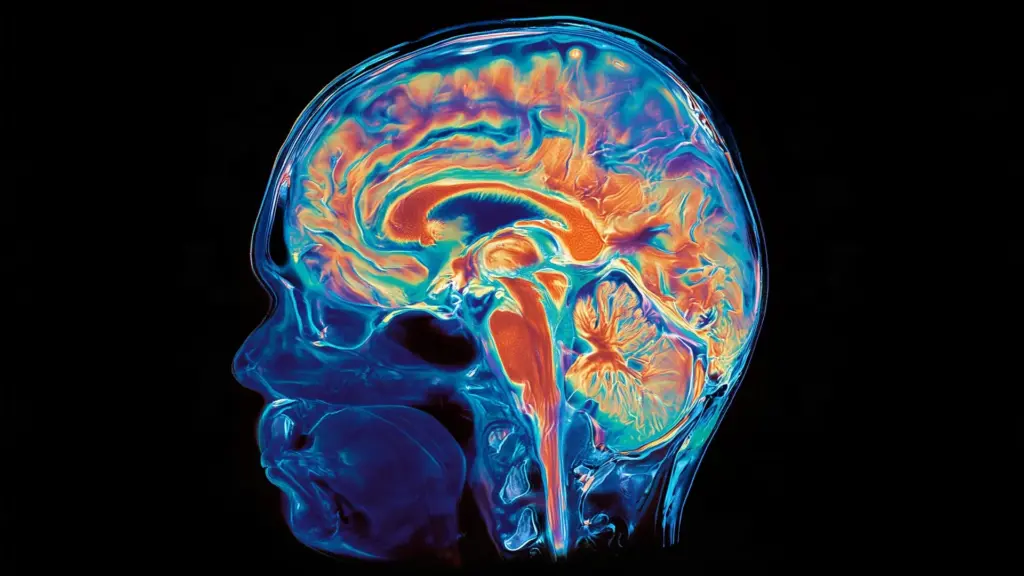
For decades, the central dogma of developmental neurobiology has held that the human brain is a monolithic entity—a single, unified organ arising from a common pool of progenitor cells. Under this established model, the forebrain, midbrain, and hindbrain were thought to trace their lineage back to the same developmental starting point, evolving in tandem as the brain grew more complex. However, groundbreaking new research led by scientists at Stanford Medicine is fundamentally challenging this assumption, revealing that what we perceive as a single brain is, in reality, a biological hybrid. The study suggests that the brain is constructed from two entirely distinct nervous systems that evolved separately over hundreds of millions of years before converging into the organ we recognize today.
The implications of this discovery are profound, potentially rewriting textbooks on human development while providing a long-sought solution to a persistent mystery in medical research: why scientists have historically struggled to cultivate certain brain cells in laboratory settings. By identifying the separate origins of these two brain systems, researchers have unlocked a new pathway for investigating and potentially treating neurodegenerative diseases, including spinal muscular atrophy (SMA) and amyotrophic lateral sclerosis (ALS).
The Architecture of a Dual-Origin Brain
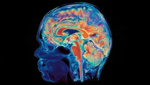
The findings, published on September 18 in Nature Neuroscience, offer a radical departure from the long-standing consensus. The research team, led by senior author Kyle Loh, PhD, an associate professor of developmental biology at Stanford, found that the brain appears to be a composite of two ancient, independent systems. One system gives rise to the structures that govern the body’s most vital, involuntary functions—such as the regulation of heartbeat, respiration, sleeping patterns, and the hunger-satiety balance. This is the hindbrain, or brain stem. The second system produces the regions responsible for the hallmarks of human consciousness: abstract reasoning, complex language, mathematics, and the ability to reflect on our own existence.

This distinction is not merely academic. For researchers attempting to model brain diseases in a petri dish, the inability to effectively grow hindbrain neurons has been a persistent roadblock. "We’ve shown for the first time that the front of the brain arises from a totally different progenitor cell than the back of the brain," said Loh. "Our discovery means that we can now grow neurons from the back of the brain—the hindbrain—in a petri dish and study their functions."
The research was spearheaded by graduate students Carolyn Dundes and Rayyan Jokhai, who serve as co-first authors on the study. Their work clarifies that the brain’s tripartite structure—divided into the forebrain, midbrain, and hindbrain—masks a much deeper developmental divide. While the forebrain handles the cognitive heavy lifting, the hindbrain sits at the base of the skull, managing the automatic processes that keep us alive. Furthermore, hindbrain neurons are responsible for the intricate control of muscles in the face, tongue, and throat, which are essential for speaking and swallowing.
The struggle to replicate these neurons in a laboratory setting has hindered progress in understanding diseases like ALS, often known as Lou Gehrig’s disease, and SMA. In both conditions, specific hindbrain neurons begin to fail. As these cells degrade, patients may lose the ability to swallow, leading to a high risk of life-threatening pneumonia when food or liquids are inhaled into the lungs. Eventually, the loss of control over respiratory functions can become fatal. Because researchers were previously unable to grow these specific neurons, they were essentially working in the dark, unable to observe the early-stage progression of these disorders in a controlled environment.
A Split Rooted in Embryonic Development
The breakthrough occurred when the researchers shifted their focus to the earliest stages of embryonic development, specifically the period known as gastrulation, when the body’s basic structure is first established. It was here that Dundes and Jokhai discovered that the hindbrain does not emerge as a secondary, later branch of the developmental pathway that produces the forebrain and midbrain. Instead, it follows a completely independent trajectory from the very beginning, developing in parallel with the other regions.
To map this process, the team studied developing mouse embryos, successfully identifying two distinct populations of brain progenitor cells. One population, characterized by the expression of the gene Otx2, is destined to form the forebrain and midbrain. A second, entirely separate population expresses the gene Gbx2 and is exclusively responsible for the hindbrain. Crucially, the researchers observed that these two populations remain distinct and do not overlap, even at the earliest developmental stages examined.
The distinction was further corroborated by analyzing chromatin, the complex of DNA and proteins that packages genetic material within the cell. By acting as a gatekeeper, chromatin determines which genes remain accessible and which are silenced. The team found that the anterior neural ectoderm (the future forebrain and midbrain) and the posterior neural ectoderm (the future hindbrain) possessed fundamentally different chromatin configurations. These epigenetic differences effectively committed the cells to separate developmental fates long before the physical structures of the brain were even formed.
"Previous attempts to make hindbrain neurons likely tried to coax forebrain and midbrain progenitors into hindbrain cells, which our study shows is not possible," explained Jokhai. This realization solves a riddle that has frustrated stem cell biologists for years: researchers had been attempting to force progenitor cells to adopt a fate they were never biologically capable of fulfilling. "In stem cell biology, people are always fixated with creating the end cell type, like the neuron," Jokhai noted. "But it’s important to begin at the earliest stages of embryonic development. Our careful attention to that early time point allowed us to find this fundamental split in brain development."
Harnessing New Knowledge for Disease Research
Armed with the knowledge that the hindbrain follows a separate evolutionary and developmental path, the researchers successfully achieved a milestone in stem cell biology: they guided human pluripotent stem cells—cells capable of becoming any tissue type in the body—into becoming fully functional hindbrain motor neurons. These laboratory-grown cells displayed the hallmark behaviors of authentic hindbrain neurons, including the production of electrical signals known as action potentials and the synthesis of proteins required for the control of facial and swallowing muscles.
The evolutionary scope of this finding is equally striking. By looking back over 550 million years of history, the team found evidence of this two-origin arrangement in species as diverse as chickens, zebrafish, and acorn worms. Even more remarkably, jellyfish—which diverged from the human evolutionary lineage roughly 600 to 700 million years ago—possess two nervous systems situated at opposite ends of their bodies.
These findings support the hypothesis that the modern vertebrate brain was formed when evolution physically merged two preexisting, independent neural systems. "Our research suggests that evolution took two existing neural systems and pushed them together spatially," said Loh. "Having the brain as one organ would probably be more efficient, but we rely on this primordial way to make the brain as two separate pieces."
For Jokhai, the findings were both surprising and enlightening. "I was surprised at our findings because the word ‘brain’ implies a contiguous organ that likely has a singular origin," he said. "But even 500 million years ago, there were these separate neural systems, which now almost operate as one, which is very cool."
Looking ahead, this discovery offers a new, high-precision tool for medical research. Beyond ALS and SMA, the hindbrain is central to the regulation of metabolism and hunger, processes that are increasingly relevant to the development of new weight-loss medications. By providing a reliable, repeatable model for growing hindbrain neurons, this research provides a vital window into the mechanics of the brain stem. The Stanford team now plans to investigate the developmental origins of the spinal cord and further explore how these distinct neural systems are disrupted by disease, opening an exciting new frontier in the quest for regenerative therapies.
The study involved contributions from researchers at the California Institute of Technology and the University of California, San Francisco, and received broad support from organizations including the National Institutes of Health, the National Science Foundation, the California Institute for Regenerative Medicine, the Spinal Muscular Atrophy Foundation, and the Howard Hughes Medical Institute, among several other philanthropic and research centers.