








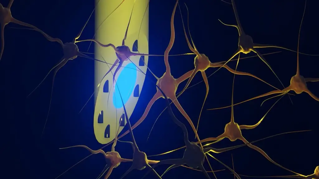
A revolutionary leap in neurotechnology has emerged from an international collaboration between the Technical University of Denmark (DTU), the University of Copenhagen, and University College London. Researchers have successfully developed a cutting-edge, needle-thin brain implant capable of unprecedented multi-modal interaction with neural tissue. Known as the microfluidic Axialtrode (mAxialtrode), this device promises to transform how scientists investigate the complex architecture of the brain, potentially paving the way for future clinical interventions in debilitating neurological conditions, including epilepsy.
The findings, which were recently published in the prestigious journal Advanced Science, detail a device that functions as a Swiss Army knife for neuroscientists. By integrating multiple functional points along the length of a single, flexible implant, the mAxialtrode allows for the simultaneous recording of electrical neural activity and the targeted delivery of pharmacological agents across various layers of the brain.
A Multifunction Tool for Brain Research
For the present, the mAxialtrode is being positioned primarily as a transformative research instrument. The complexity of the human brain—and the brains of mammalian models—relies on intricate signaling pathways that span multiple layers and anatomical structures. Current research methodologies often struggle to capture the full picture of these interactions, as they are frequently limited to observing localized activity.
With this new technology, scientists gain the ability to investigate how signals propagate through different layers of the brain during fundamental processes such as memory formation, complex decision-making, and the erratic neural firing patterns characteristic of epilepsy. By providing a longitudinal view of these circuits, the device offers a level of spatial resolution that was previously difficult to achieve without utilizing multiple, invasive probes.
Over the longer term, the research team envisions a future where the mAxialtrode transitions from the laboratory to the clinic. The potential therapeutic applications are significant; the device could be used to deliver medication to precise, deep-seated locations while simultaneously modulating neural behavior through electrical or light-based stimulation. This "closed-loop" approach, where a device monitors, stimulates, and treats in real-time, represents a major goal in modern neuro-engineering.
Postdoc Kunyang Sui, who conceptualized the mAxialtrode alongside Associate Professor Christos Markos, emphasizes that the integration of multiple capabilities into a single implant is the device’s most defining advantage. "By combining recording, light stimulation, and drug delivery into one structure, we can perform significantly more precise experiments," Sui explains. "This consolidation also reduces the physical burden on the brain, as it minimizes the need for researchers to insert multiple, separate devices to perform different tasks."
Overcoming the Limitations of Traditional Implants
A primary challenge in neuro-implant design has always been the compatibility between rigid hardware and delicate biological tissue. Traditional brain implants are frequently manufactured from silicon or other hard, metallic materials. While these materials are effective for recording electrical signals, their stiffness often leads to chronic irritation and secondary inflammatory reactions. This inflammatory response can degrade the quality of recorded signals over time and potentially cause long-term damage to the very tissue researchers aim to study.
The mAxialtrode marks a significant departure from these conventions. "The new implant is made of soft, plastic-like optical fibers," says Sui. "It also features a specially angled tip that makes it smaller and reduces the trauma caused to the brain during insertion." Because the device is highly flexible, it moves in tandem with the brain tissue, reducing the mechanical strain that typically triggers glial scarring—the body’s defensive process of walling off foreign, rigid objects.
This shift toward flexible electronics is a growing trend in the field of neural interfaces, but the mAxialtrode elevates the concept by incorporating fluidic capabilities into a structure thinner than a human hair. Despite its promising design, the team remains cautious. Sui is clear that the technology is currently in its nascent stages and remains far from routine clinical application. Extensive validation, rigorous long-term safety testing, and the navigation of complex regulatory landscapes are essential steps that must occur before the device could ever be considered for use in human patients.
Moving Beyond Conventional Optical Fibers
To understand the innovation behind the mAxialtrode, it is necessary to consider the standard tools currently used in neuroscience. For years, researchers have relied on flat-ended optical fibers to probe the brain. These thin filaments, typically made from glass or plastic, are the workhorses of optogenetics—a revolutionary technique that allows scientists to selectively activate or inhibit specific neurons using pulses of light.
However, these conventional fibers suffer from a significant structural limitation: they are essentially "end-fire" devices. They can only interact with brain tissue at the distal tip—the very end of the fiber. This means that a researcher is restricted to monitoring or stimulating only one specific location. In the context of brain function, where signals often cascade through different layers and deeper structures simultaneously, this "single-point" limitation forces scientists to make compromises in their data collection. If a study requires mapping a circuit that spans three millimeters of vertical depth, a traditional fiber would have to be repositioned multiple times, increasing the risk of tissue damage and reducing the reliability of the resulting data.
How the New Brain Implant Works
The engineering process behind the mAxialtrode is a testament to precision manufacturing. The fiber begins its life as a much larger polymer rod. Through a process involving carefully controlled heating and drawing, the material is thinned down into a fine, needle-like strand, not unlike the way fine sugar strands are drawn in culinary arts, albeit with sub-micron levels of precision.
The internal architecture of the resulting fiber is remarkably complex. At its heart lies a light-conducting core designed for optical stimulation. Encircling this central core are eight microscopic channels. These channels serve a dual purpose: they can be used to transport liquids, such as neurotransmitters or therapeutic drugs, directly to the desired depth, or they can be used to house ultra-thin metal wires. These wires function as high-resolution electrodes, capable of picking up the delicate electrical chatter of individual neurons.
The finished device measures less than half a millimeter in diameter. This scale allows for minimal displacement of brain matter upon insertion, further protecting the integrity of the neural environment. Its mechanical flexibility is the final piece of the puzzle, allowing the implant to remain stable within the intracranial environment without the rigid pressure associated with traditional silicon probes.
Tested in Living Mice
The research team moved from theoretical design to practical application by conducting in vivo tests in living mice. This step was crucial to demonstrate that the device could function reliably in a complex, biological system. The electrode was carefully implanted into the animals’ brains and integrated with a comprehensive control system, including external light sources, sensitive recording equipment, and precision micro-pumps for fluid delivery.
The results were compelling. The researchers successfully demonstrated the device’s ability to stimulate nerve cells using both blue and red light—a critical requirement for complex optogenetic experiments. Simultaneously, they recorded electrical activity across various depths, successfully capturing data from both the cerebral cortex and the deeper-lying hippocampus.
Perhaps most impressive was the device’s ability to inject different substances at precise, separate depths. The researchers were able to deliver fluids at points spaced nearly three millimeters apart along a single fiber. Throughout these demanding procedures, the mice exhibited no obvious signs of discomfort or impairment, carrying the lightweight fiber as they moved freely.
Potential Applications in Epilepsy and Neuroscience
The success of these in vivo experiments was made possible through the interdisciplinary synergy of the research team. Associate Professor Rune W. Berg of the University of Copenhagen and Associate Professor Rob C. Wykes from University College London provided vital expertise in neurophysiology and the analysis of neural circuits, particularly those modeled in epilepsy research. By aligning the engineering capabilities of DTU with the clinical and physiological knowledge of their collaborators, the team was able to validate the device’s utility in real-world neuroscience research.
As the team moves forward, they are currently working to secure patents for the technology, ensuring that the intellectual property behind the mAxialtrode is protected. Meanwhile, the scientists are beginning the arduous process of defining the roadmap required to transition the device toward clinical testing. While the path to human use is long and fraught with technical and regulatory hurdles, the development of the mAxialtrode provides a new, highly precise tool that could eventually change the way we treat some of the most complex and persistent neurological conditions known to medicine.