







Deep within the brainstem, tucked away in a region known as the locus coeruleus (LC), lies a tiny cluster of nerve cells that exerts a disproportionate influence over our daily existence. The name itself, derived from the Latin for "blue place," hints at its unique biological character, yet for decades, neuroscientists have struggled to fully map the extent of its influence. While the LC accounts for only a minuscule fraction of the brain’s total neuronal population, it serves as the primary engine for producing norepinephrine (NE), a potent chemical messenger—or neuromodulator—that governs our attention, learning capacity, heart rate, and our primal responses to stress and excitement.
For generations, the prevailing scientific consensus held that the LC functioned much like a broadcast loudspeaker, a singular, monolithic system that indiscriminately doused the brain in norepinephrine. Under this "foghorn" model, it was assumed that whenever the LC fired, it sent a uniform signal to every corner of the central nervous system, eliciting a broad, generalized state of arousal. However, a landmark study published in the journal Nature has fundamentally challenged this long-standing assumption, revealing a far more sophisticated, nuanced, and structurally precise internal architecture than previously imagined.
A Hidden Brain Network That Sends Precise Messages
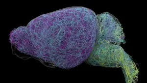
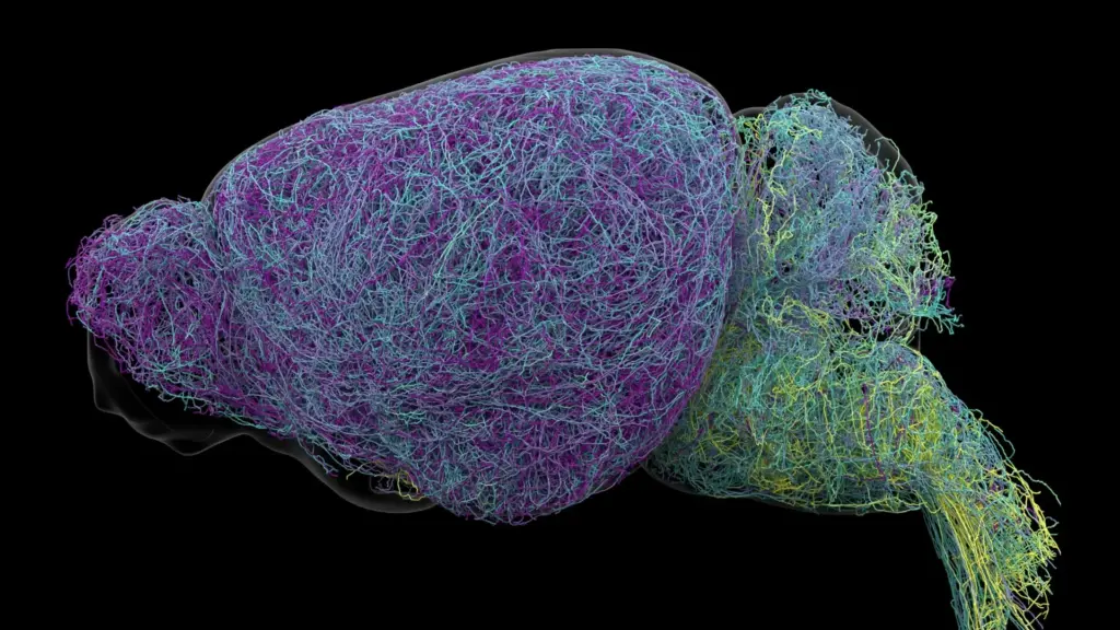
Through a multi-modal investigation involving advanced brain imaging, high-fidelity neural recordings, rigorous genetic profiling, and complex behavioral experiments in mice, researchers at the Allen Institute for Brain Science have dismantled the idea of the LC as a uniform broadcaster. Instead, the team discovered that the LC communicates through a series of highly distinct, specialized pathways. Rather than releasing one sweeping, global signal, the LC acts as a sophisticated postal network, delivering specific instructions to targeted destinations.

By meticulously tracing the physical connections of these neurons, the research team found that distinct subpopulations of cells are wired to specific regions of the nervous system. This discovery shifts our perception of the LC from a simple "volume knob" for the brain to an organized switchboard. The researchers observed that the anatomical location of a neuron within the LC determines its target: neurons situated in the upper, or dorsal, portion of the structure primarily extend their reach toward the cerebral cortex and the forebrain. Conversely, those nestled in the lower, or ventral, portion are tasked with signaling to the brainstem and the spinal cord.
"The findings suggest the brain’s norepinephrine system is far more like a targeted postal network than a foghorn," remarked Karel Svoboda, director of Neural Dynamics at the Allen Institute and a co-author of the study. This organizational logic allows the brain to modulate separate functions—such as high-level cognitive deliberation versus autonomic, life-sustaining reflexes—with granular precision.
Why the Discovery Could Matter for Brain Disorders
The implications of this structural discovery extend well beyond basic biology; they offer a potential roadmap for the future of clinical neurology and psychiatry. Norepinephrine is widely recognized as a cornerstone of mood and attention regulation. Consequently, it has long been a primary target for pharmacological interventions, including many common medications prescribed for depression, anxiety, and Attention Deficit Hyperactivity Disorder (ADHD).
However, current pharmaceutical approaches are often blunt instruments. Because existing drugs influence the entire norepinephrine system, they frequently impact multiple brain circuits simultaneously. While a medication might successfully alleviate symptoms of anxiety, it may simultaneously trigger unwanted side effects elsewhere in the brain or body due to this lack of selectivity. The new, detailed map of the LC’s specialized pathways provides a tantalizing possibility: if scientists can understand which specific circuits govern particular symptoms, they might eventually develop next-generation therapies that act selectively, "tuning" only the circuits involved in a specific disorder while leaving the rest of the brain’s delicate chemical balance undisturbed.
The urgency of this research is further underscored by the role of the locus coeruleus in neurodegeneration. LC neurons are among the very first brain cells to show signs of deterioration in the progression of Alzheimer’s disease. By mapping the precise connectivity of these cells, researchers hope to gain a clearer understanding of why these particular neurons are so vulnerable, potentially opening new windows into early intervention strategies. This research was made possible through support from the National Institutes of Health’s Brain Research Through Advancing Innovative Neurotechnologies (BRAIN) Initiative, which seeks to revolutionize our understanding of the human brain.
Scientists Identify Brain Cells Involved in Learning
Beyond the structural mapping, the researchers uncovered functional distinctions that suggest a deep division of labor within the LC. By observing the activity of these neurons during behavioral tasks, the team identified a clear separation between the "dorsal" and "ventral" roles.
When mice were tasked with decision-making—specifically the process of updating their choices following an unfavorable or unexpected result—neurons in the dorsal LC showed a marked increase in activity. This suggests that these cells are essential components of the brain’s learning machinery, helping the animal process feedback and adapt its behavior accordingly.
In contrast, the ventral LC neurons exhibited a different behavioral signature. Their activity spiked shortly before the mice chose to ignore cues that would have otherwise indicated the availability of a reward. This suggests that the ventral region acts as a gatekeeper, modulating an animal’s motivation and determining whether it chooses to engage with its environment or remain passive. As Svoboda explained, "What emerged was a clear map: neurons in the dorsal LC that send signals upward to the cortex are involved in learning, whereas neurons in the ventral region projecting downward to the brainstem and spinal cord govern whether animals engage with their environment at all."
Crucially, the study demonstrated that these functional and anatomical differences are not mere coincidences of location; they are supported by distinct gene expression patterns. The dorsal and ventral neurons are biologically unique, reflecting their specialized roles through different molecular signatures.
Researchers Discover a Record-Breaking Neuron
The sheer scale of the Allen Institute’s investigation is unprecedented. To map this network, scientists conducted whole-brain imaging on nearly 35,000 individual neurons and analyzed the genetic profiles of almost 400,000 cells. Perhaps most strikingly, they successfully reconstructed the complete, three-dimensional structures of several exceptionally complex nerve cells.
During this meticulous reconstruction, the team encountered a biological anomaly: LC neurons possess axons of extraordinary length. Axons are the long, thread-like projections that carry electrical impulses away from the cell body to distant synapses. The researchers found that, on average, these LC axons stretched approximately 35 centimeters—an impressive distance for a mouse. However, one specific neuron stood out, with a total reconstructed length of 70.32 centimeters. This represents the longest single neuron ever recorded in a mouse, a feat of biological engineering that allows a single cell to exert influence across vast swaths of the brain.
"This neuron, like many others that we studied, supplies NE to a very large volume of the cerebral cortex," explained Jeremiah Cohen, a scientist at the Allen Institute and study co-author. "For the brain, this is highly unusual. Most neurons are more specific in their targets." Yet, even this massive, record-breaking neuron maintains a sense of purpose. Despite its reach, it does not release norepinephrine everywhere; it remains highly selective, pointedly ignoring areas like the cerebellum and the spinal cord. This reinforces the core thesis of the study: the LC is not a chaotic broadcaster but a masterfully organized signaling system.
A New Understanding of How the Brain Learns
The study also provides a new lens through which to view the interplay between different chemical messenger systems. The researchers noted intriguing similarities between the LC’s norepinephrine network and the brain’s dopamine system, which is centered in the basal ganglia and is vital for habit formation. Scientists now hypothesize that dopamine and norepinephrine may operate in tandem, forming a coordinated team that supports the brain’s ability to acquire and process complex, abstract concepts simultaneously.
By revealing the intricate wiring, the specialized functional roles, and the surprising physical architecture of the locus coeruleus, this research provides a vastly more detailed picture of how the brain manages the constant influx of sensory information and transforms it into knowledge. While these findings were derived from mouse models, they provide a foundational framework that will likely inform future studies of the human brain. The transition from viewing the LC as a simple "foghorn" to understanding it as a sophisticated, targeted, and highly organized communication hub marks a significant milestone in neurobiology, offering a clearer path toward decoding the biological basis of our most complex cognitive behaviors.