








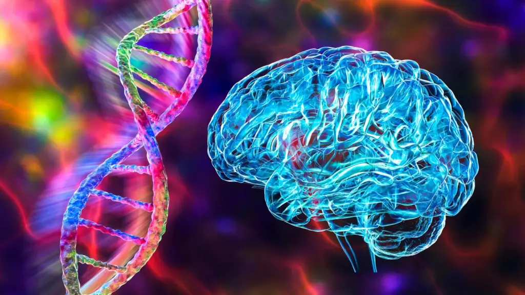
For decades, the medical community has grappled with a confounding mystery: why do some individuals, despite carrying the most significant genetic risk factor for Alzheimer’s disease, remain cognitively sharp and symptom-free well into their 80s and beyond? This genetic resilience has long been a focal point for researchers seeking to unlock the secrets of neuroprotection. Now, a team of scientists at Columbia University has identified a specific biological mechanism that appears to provide this natural defense, potentially offering a transformative roadmap for the future of Alzheimer’s treatment.
The discovery centers on the interplay between the well-known APOE ε4 gene variant—the strongest genetic risk factor for the disease—and a rare, protective mutation in the fibronectin (FN1) gene. According to findings published in the journal Nature Aging, this rare mutation acts as a biological shield, safeguarding the brain’s delicate blood vessels from damage that typically leads to cognitive decline.
Unlocking a Protective Mechanism
The research builds upon a critical study published by the Columbia team two years ago, which first identified that certain individuals carrying the high-risk APOE ε4 variant also possessed a protective mutation in the fibronectin gene. Fibronectin is a protein essential for structural support throughout the human body. However, in the context of the APOE ε4 variant, the body often produces excessive amounts of this protein. When this surplus accumulates around the brain’s blood vessels, it can compromise the integrity of the blood-brain barrier, the critical system that regulates the movement of substances between the bloodstream and the brain.
The newly published study bridges the gap between genetic association and biological causality. By identifying exactly how excess fibronectin causes harm, the researchers have pinpointed potential therapeutic targets that could mimic the protective effects of the rare FN1 mutation.
"This new study takes us from a genetic clue to a disease mechanism," says Caghan Kizil, the study’s corresponding author and professor of neurological sciences at Columbia University’s Vagelos College of Physicians and Surgeons. "We knew that changes in fibronectin could protect against Alzheimer’s, but we didn’t know why excess fibronectin was harmful in the first place. This study gives us that detailed mechanism—and with it, ideas for how to reproduce that natural protection."
How an Alzheimer’s Risk Gene Weakens the Blood-Brain Barrier
To understand the scope of the discovery, one must first understand the function of the blood-brain barrier. This barrier is a sophisticated, protective boundary composed of tightly connected cells that wrap around the brain’s blood vessels. Its primary role is to act as a gatekeeper, carefully controlling which substances enter brain tissue while ensuring the environment is stable enough to facilitate the removal of metabolic waste.
When this barrier becomes leaky or permeable—a process often linked to aging and neurodegenerative conditions—it allows harmful substances from the bloodstream to infiltrate the brain. This intrusion can trigger inflammation and other biochemical cascades that are hallmark features of Alzheimer’s disease.
The Columbia research team employed a multi-faceted approach to investigate the link between fibronectin and this barrier failure. They utilized genetic and epigenetic analyses on human brain tissue and cerebrospinal fluid, coupled with single-cell sequencing and spatial imaging. These advanced methodologies allowed the team to pinpoint where biological changes occur at the cellular level. To validate their hypotheses, the researchers also conducted experiments using human stem-cell models, three-dimensional cultures of blood vessels, and animal models, including zebrafish and mice.
The results were consistent across all models: the presence of the APOE ε4 gene variant directly promotes the production and subsequent accumulation of fibronectin around the brain’s blood vessels. In mice engineered to express human APOE ε4, the researchers observed nearly double the levels of fibronectin compared to those carrying the more common APOE ε3 variant. Crucially, this accumulation was directly correlated with evidence that the blood-brain barrier had lost its structural integrity.
Scientists Discover How Fibronectin Causes Brain Damage
Moving beyond simple association, the researchers sought to determine if fibronectin was the primary driver of this damage. They focused their efforts on astrocytes, the specialized support cells that are vital for maintaining the blood-brain barrier. By increasing the expression of human fibronectin specifically within these cells, the team observed a direct disruption of the barrier, rendering it permeable. Conversely, when they reduced fibronectin levels, the function of the barrier improved significantly.
"The animal experiments allowed us to move beyond association," notes co-first author Prabesh Bhattarai, an associate research scientist in the Kizil lab. "When we increased fibronectin in astroglia, the barrier became permeable. When we reduced fibronectin, barrier function improved."
The team then mapped the molecular events responsible for this breakdown. They discovered that excessive fibronectin interferes with intercellular communication by activating abnormal signaling through integrins—proteins that allow cells to sense and respond to their environment—and an enzyme called focal adhesion kinase.
This signaling malfunction disrupts a network of essential growth factors, specifically VEGF, HBEGF, and IGF1. While these growth factors are typically responsible for cell development and repair, they also play a vital role in helping astrocytes and blood vessel cells coordinate their efforts to maintain a healthy, impermeable barrier. When this communication is severed, the barrier fails, leaving the brain vulnerable to the toxic processes that characterize Alzheimer’s pathology.
Co-first author Elanur Yilmaz, an associate research scientist in the Kizil lab, emphasizes the consistency of these findings. "Single-cell analysis allowed us to examine this pathway in the different cells that maintain the brain’s blood vessels. We observed the same biological pattern across experimental models and human Alzheimer’s tissue, making the mechanism especially compelling."
Evidence From Human Brains Strengthens the Alzheimer’s Connection
The laboratory findings were bolstered by an analysis of human brain samples, which provided a crucial link to clinical reality. The data showed that elevated fibronectin levels were associated with significant inflammation in astrocytes. Furthermore, genetic and gene-regulation analyses connected the FN1 gene to vascular disease and alterations involving VEGFA, a growth factor essential for maintaining the health of the brain’s vascular system.
Badri Vardarajan, associate professor of neurological science and a collaborator on the study, noted that the convergence of these independent datasets provides a robust argument for the role of fibronectin in the disease process. "The convergence of these independent human datasets strengthens the case that fibronectin is part of the APOE ε4-related vascular disease process," Vardarajan said.
A Rare Genetic Defense Could Inspire New Alzheimer’s Treatments
The implications of this study are profound, particularly given that APOE ε4 is the most significant common genetic risk factor for Alzheimer’s, affecting approximately one in five people. While not every carrier will develop the disease, the increased risk is substantial. By uncovering why some individuals remain resilient, researchers have opened a new door for preventative medicine.
The researchers have identified three potential therapeutic strategies: preventing the accumulation of harmful fibronectin, blocking the abnormal cellular signals triggered by excess fibronectin, or restoring the growth factor signaling that supports the blood-brain barrier.
The path forward, however, requires nuance. Because fibronectin is a critical protein for healing and tissue structure throughout the body, any systemic treatment must be carefully designed to target the harmful accumulation in the brain without interfering with the protein’s necessary, healthy functions elsewhere.
"By showing how fibronectin contributes to early vascular damage, this study gives us new therapeutic targets and potential ways to measure whether treatments are working," says Richard Mayeux, chair of the Department of Neurology at Columbia and a project collaborator. "Clinically, it connects a major genetic risk factor for Alzheimer’s to an early disease process that may be possible to modify."
While therapies derived from this research remain in the experimental stage, the discovery marks a significant pivot in the fight against Alzheimer’s. By shifting the focus toward mimicking natural genetic defenses and addressing early vascular damage, scientists are moving closer to a future where they can intervene before the devastating effects of the disease take hold.