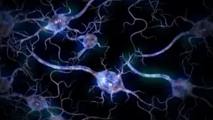









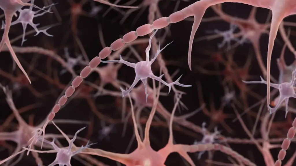
For over a century, textbooks have taught students that axons—the long, slender extensions of neurons responsible for transmitting electrical signals—are essentially smooth, uniform tubes. This classic illustration, which depicts these vital cellular cables as narrow, consistent conduits, has served as a foundational concept in neuroscience. However, a groundbreaking study from Johns Hopkins Medicine, published in Nature Neuroscience in December 2024, suggests that this long-standing mental model may be incomplete.
Researchers discovered that, rather than being perfectly smooth, many axons actually possess a repeating, "pearled" architecture. These tiny, rhythmic bulges, which the team calls "non-synaptic varicosities," are not merely structural anomalies or artifacts of tissue preparation. Instead, they appear to be a fundamental feature of normal axon anatomy, exerting a measurable influence on how electrical messages travel through the brain.
A New Anatomy of Brain Cell Signaling
Axons are the fundamental cables that connect the brain’s vast neural network, facilitating the complex signaling required for learning, memory, and motor function. Traditionally, scientific illustrations have depicted them as smooth tubes, occasionally punctuated by larger, well-known swellings called synaptic varicosities, which house the neurotransmitters necessary for signaling at junctions.

"Understanding the structure of axons is important for understanding brain cell signaling," says Shigeki Watanabe, Ph.D., an associate professor of cell biology and neuroscience at the Johns Hopkins University School of Medicine. "Axons are the cables that connect our brain tissue, enabling learning, memory, and other functions."
The existence of beaded or swollen axons is not entirely new to the scientific community. For decades, researchers have observed pronounced "beading" in dying neurons and in the context of neurodegenerative conditions, such as Parkinson’s disease. In these pathological settings, the beading is typically associated with a breakdown of the cell membrane and the internal protein framework that provides structural integrity to the axon.
What made the Johns Hopkins findings so striking was the observation of much smaller, repeating swellings in healthy axons under conditions specifically designed to preserve their native state. By distinguishing these nanoscale "non-synaptic varicosities" from the larger, damage-related swellings observed in disease, the researchers established that this pearling is a feature—not a flaw—of the healthy brain.
The Influence of Membrane Physics
The research team’s journey to this discovery began with curious observations of repeating pearls in the axons of worms. Watanabe, in consultation with Swiss scientist Graham Knott, Ph.D., initially hypothesized that the cell’s internal protein skeleton might be responsible for shaping these structures. However, when the team disrupted this internal framework, the pearling remained intact, suggesting that the explanation lay elsewhere.
To solve the puzzle, Watanabe and graduate student Jacqueline Griswold collaborated with theoretical biophysicist Padmini Rangamani, Ph.D., a professor of pharmacology at the University of California San Diego. Together, they investigated whether the physical properties of the surrounding cell membrane could explain the formation of these pearls.
Studying structures roughly 100 times thinner than a human hair required extreme precision. The team employed high-pressure freezing electron microscopy to preserve the axons in their natural state. Standard imaging techniques often involve chemical fixation and dehydration, which can inadvertently alter delicate cellular architectures. By "freezing" the tissue rapidly, the researchers were able to capture a snapshot of the axon that closely resembled its appearance in a living state.
"To see nanoscale structures with standard electron microscopy, we fix and dehydrate the tissues, but freezing them retains their shape—similar to freezing a grape rather than dehydrating it into a raisin," Watanabe explained.
Across tens of thousands of images of mouse neurons—including those grown in the lab, neurons from embryos, and those from adult mice—the pearled pattern was consistent. The team also confirmed these findings through high-resolution imaging of living neurons, solidifying the evidence that the pearling was not a byproduct of the freezing process.
Mathematical Models and Membrane Mechanics
The researchers developed mathematical models of the axonal membrane to test their hypothesis. They found that relatively simple mechanical forces could replicate the pearls without requiring a rigid internal mold. By adjusting variables in their models, they could manipulate the size of the bulges.
Experiments in the laboratory supported these models. When the researchers increased the sugar concentration in the surrounding environment, the swellings decreased in size. Similarly, when they adjusted membrane tension, the size of the pearls shifted accordingly.
The composition of the membrane itself—specifically cholesterol content—also played a critical role. When cholesterol was removed, the membrane became more fluid and less stiff. This modification altered the pearled structure and, crucially, slowed the speed of electrical signaling through the axon.
"A wider space in the axons allows ions [chemical particles] to pass through more quickly and avoid traffic jams," Watanabe said. However, he noted that the relationship is nuanced; both the dimensions of the swellings and the narrow connecting segments are essential factors in determining how efficiently signals move.
Activity-Dependent Structural Changes
Perhaps most intriguing is the discovery that these axons are not fixed, immutable cables. The researchers found that neural activity itself can reshape the membrane. After high-frequency electrical stimulation, the pearl-like regions increased in length and width—an effect that persisted for at least 30 minutes.
This structural plasticity appeared to have functional consequences. Following stimulation, electrical signals through the affected axons slowed, an effect that lasted for at least an hour. In cells where cholesterol had been removed to alter membrane stiffness, these structural changes were blunted, and the significant slowing of signals was not observed. These findings suggest that the physical architecture of the axon is dynamic, constantly adjusting its shape in response to activity to modulate the flow of information.
From Mice to Humans
The significance of these findings extends beyond mouse models. Following the announcement of the initial results, researchers sought to determine if this architecture exists in human tissue. A subsequent study, published in Neuron in February 2026, confirmed the presence of pearled axons in human cortical tissue obtained from patients undergoing epilepsy surgery.
This research, led by Chelsy R. Eddings and colleagues—including Watanabe—utilized a combination of electrical stimulation and rapid freezing to capture membrane dynamics in human brain slices. While these findings bridge the gap between rodent and human neuroscience, they also underscore the complexity of the brain. The researchers emphasized that these observations do not mean every axon possesses the same shape, nor do they suggest that pearling has a uniform impact on signaling across all regions of the human brain. Much of this work has focused on membrane recycling at synapses, leaving further questions about the broader distribution of these structures.
The Future of Axonal Research
The relationship between structural integrity and neuronal function remains a primary focus for the field. A multi-institutional project, supported by the National Institute of Mental Health (NIMH), is currently expanding on this work through advanced computational modeling and further experimentation. The goal is to better understand how the physical properties of a neuron and its environment dictate the behavior of its axons.
Meanwhile, other researchers are developing innovative tools to investigate axonal dimensions. In a study published in PLOS Biology in July 2026, researchers at the University of Edinburgh utilized automated imaging of living zebrafish to screen hundreds of chemical compounds, identifying several that could influence axon width. While this work does not directly replicate the pearling study, it provides a powerful new toolkit for understanding the regulation of axon morphology.
For the Johns Hopkins team, the most pressing question remains the relationship between these normal, functional pearls and the pathological beading seen in neurodegenerative diseases. By clearly defining what constitutes "normal" structural variance, scientists hope to establish a diagnostic baseline. Distinguishing between the adaptive, dynamic changes of a healthy, functioning brain and the irreversible, damaging beading of a system in decline could be a vital step toward developing new therapeutic interventions for neurological disorders.
As the scientific community continues to move beyond the "smooth tube" dogma, the realization that axons are complex, dynamic structures capable of modulating their own signaling environment opens a new frontier in our understanding of how the brain processes information. Far from being simple wires, these axons are active participants in the symphony of neural communication.