







Deep within the brainstem, nestled in a region that translates from Latin to the "blue place," lies a tiny, unassuming cluster of nerve cells known as the locus coeruleus (LC). For decades, this region has been a subject of intense fascination for neuroscientists. Despite occupying a mere fraction of the brain’s total volume, the LC acts as a primary manufacturing hub for norepinephrine (NE), a potent chemical messenger—or neurotransmitter—that governs everything from our attention span and learning capabilities to heart rate and our complex physiological responses to stress and excitement.
For a long time, the prevailing scientific consensus was that the locus coeruleus functioned much like a broadcast tower or a loudspeaker. It was widely assumed that when the LC fired, it sent a singular, undifferentiated signal across the brain, washing the cortex and other regions in norepinephrine to heighten general arousal. However, a groundbreaking study recently published in the journal Nature has fundamentally dismantled this long-standing theory. By leveraging advanced brain imaging, precise neural activity recordings, cutting-edge genetic techniques, and sophisticated behavioral experiments in mouse models, researchers at the Allen Institute for Brain Science have discovered that the LC is not a monolithic broadcaster. Instead, it operates through a highly complex and organized set of distinct pathways, delivering specific signals to targeted destinations with remarkable spatial precision.
A Hidden Brain Network That Sends Precise Messages
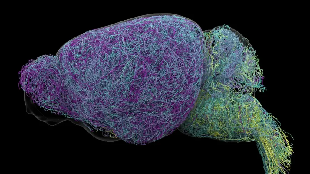
The research team set out to map the architectural connectivity of the locus coeruleus, tracing the paths of its neurons to see exactly where they terminate. What they uncovered was a far more nuanced communication network than the "foghorn" model previously suggested. The LC does not simply spray norepinephrine into the void; it directs the chemical to specific, localized regions of the nervous system.
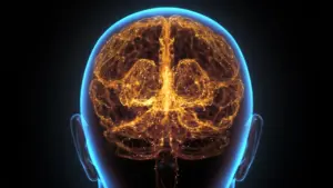
The structural map revealed a clear division of labor based on anatomical location. Neurons residing in the upper, or dorsal, portion of the locus coeruleus primarily forge connections with the cerebral cortex and the forebrain—areas of the brain associated with higher-order cognitive functions. Conversely, cells situated in the lower, or ventral, portion of the structure predominantly send their projections toward the brainstem and the spinal cord. This spatial arrangement allows different segments of this tiny nucleus to exert independent influence over vastly different bodily and mental functions.
"The findings suggest the brain’s norepinephrine system is far more like a targeted postal network than a foghorn," said Karel Svoboda, director of Neural Dynamics at the Allen Institute and a co-author of the study. By viewing the system as a specialized delivery service rather than a broadcast system, researchers can now begin to explain how the brain manages to regulate such a wide array of disparate tasks simultaneously.
Why the Discovery Could Matter for Brain Disorders
The implications of this discovery extend well beyond basic neurobiology. Because norepinephrine is central to the regulation of attention, mood, and physiological arousal, it is a primary target for a wide spectrum of psychiatric and neurological medications, including those used to manage depression, anxiety, and Attention Deficit Hyperactivity Disorder (ADHD). Furthermore, the locus coeruleus is historically significant in the study of neurodegeneration; its neurons are among the very first in the brain to succumb to the ravages of Alzheimer’s disease.
Current therapeutic approaches that aim to modulate norepinephrine levels often suffer from a lack of precision. Because these drugs typically influence multiple brain circuits at once, they can provide relief for specific symptoms while simultaneously triggering unwanted side effects in other regions of the brain. This "sledgehammer" approach is a direct result of the previous understanding that the LC was a uniform, generalized system.
With this new, granular map of the LC’s specialized pathways, scientists may be able to refine the development of next-generation therapies. By targeting the specific circuits involved in particular symptoms—rather than flooding the entire system with neurochemical agents—researchers hope to create medications that are both more effective and better tolerated. This research, which was supported by the National Institutes of Health’s Brain Research Through Advancing Innovative Neurotechnologies (BRAIN) Initiative, provides the structural foundation required for this new era of precision neuropsychiatry.
Scientists Identify Brain Cells Involved in Learning
Beyond structural organization, the study also uncovered distinct functional specializations among the LC neurons. By observing the behavior of mice during various tasks, the researchers were able to correlate specific neural activity with concrete learning and behavioral outcomes.
The neurons in the dorsal LC—those connecting to the cortex—showed a marked increase in activity specifically during learning tasks. When the mice were engaged in decision-making and needed to adjust their strategies following an unfavorable outcome, these dorsal neurons surged in activity, suggesting that they are critical for cognitive flexibility and error correction.
In stark contrast, neurons in the ventral LC displayed a completely different behavioral profile. Their activity levels spiked shortly before the mice ignored cues that a reward was available, indicating that this population of neurons plays a role in regulating environmental engagement. "What emerged was a clear map: neurons in the dorsal LC that send signals upward to the cortex are involved in learning, whereas neurons in the ventral region projecting downward to the brainstem and spinal cord govern whether animals engage with their environment at all," Svoboda explained.
Crucially, the research team discovered that these functional differences were not merely a matter of geography; they were deeply rooted in the cells’ molecular identity. The anatomical differences were mirrored by distinct gene expression patterns, confirming that the two groups of neurons are genetically distinct populations, each hard-wired for a specific physiological role.
Researchers Discover a Record-Breaking Neuron
The scale of the Allen Institute study was immense, involving the examination of nearly 35,000 neurons through whole-brain imaging and the genetic profiling of approximately 400,000 individual cells. During this meticulous reconstruction of neural structures, the team encountered a surprising find: the locus coeruleus contains neurons with axons of extraordinary length.
Axons are the long, slender projections that carry electrical and chemical signals away from the neuron’s cell body to distant targets. In the case of the LC neurons studied, these axons measured an average of 35 centimeters. However, one specific neuron reconstructed by the researchers reached a staggering 70.32 centimeters in length, earning the title of one of the longest single neurons ever documented in a mouse.
"This neuron, like many others that we studied, supplies norepinephrine to a very large volume of the cerebral cortex. For the brain, this is highly unusual," said Jeremiah Cohen, a scientist at the Allen Institute and study co-author. "Most neurons are more specific in their targets." Despite the vast reach of these axons, the team emphasized that they do not simply distribute signals haphazardly. The connections remain highly selective, sparing regions like the cerebellum and brainstem. This reinforces the central takeaway of the study: even in the case of neurons with massive, expansive reach, the brain maintains a rigorous level of structural and functional organization.
A New Understanding of How the Brain Learns
The study also shed light on how the norepinephrine system might interact with other key chemical signaling pathways, most notably dopamine. Dopamine is widely recognized for its role in the basal ganglia, a brain region fundamental to habit formation and reward processing. Scientists have long suspected that norepinephrine and dopamine work in concert to facilitate learning. The new findings suggest that these two systems may interact to allow the brain to acquire and process multiple abstract concepts simultaneously.
By providing a more detailed, map-like picture of the locus coeruleus—its intricate connections, specialized genetic functions, and remarkable physical structure—this research offers a profound leap forward in our understanding of how the brain communicates and learns. While these findings are derived from mouse models, they serve as a powerful blueprint for future human research. As scientists continue to explore these specific neural circuits, the dream of designing targeted, highly effective treatments for neurological and mental health disorders moves significantly closer to reality.