






Deep within the brainstem lies a minuscule cluster of nerve cells that wields an influence far exceeding its modest size. Known as the locus coeruleus (LC)—a name derived from the Latin for "blue place" due to its distinct pigmentation—this region has long been viewed by neuroscientists as the brain’s primary broadcast station. Despite containing only a small fraction of the brain’s total neuronal population, the LC serves as the body’s principal manufacturer of norepinephrine (NE), a vital chemical messenger that orchestrates a wide array of physiological and psychological processes, from heart rate and stress responses to the more complex domains of attention and learning.
For decades, the prevailing scientific consensus characterized the locus coeruleus as a "loudspeaker" for the brain. The assumption was that the LC functioned as a monolithic entity, sending a generalized, uniform signal across the entire organ to heighten arousal or alertness in response to external stimuli. However, a landmark study published in the journal Nature has fundamentally dismantled this long-standing theory. Researchers at the Allen Institute, utilizing a combination of high-resolution brain imaging, precise neural activity recordings, advanced genetic techniques, and behavioral experiments in mice, have discovered that the LC is far more sophisticated than once thought. Rather than acting as a broadcast system, the LC operates through a highly organized set of distinct, specialized pathways, delivering precise messages to specific destinations based on the nature of the experience.
A Hidden Brain Network That Sends Precise Messages
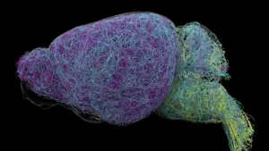
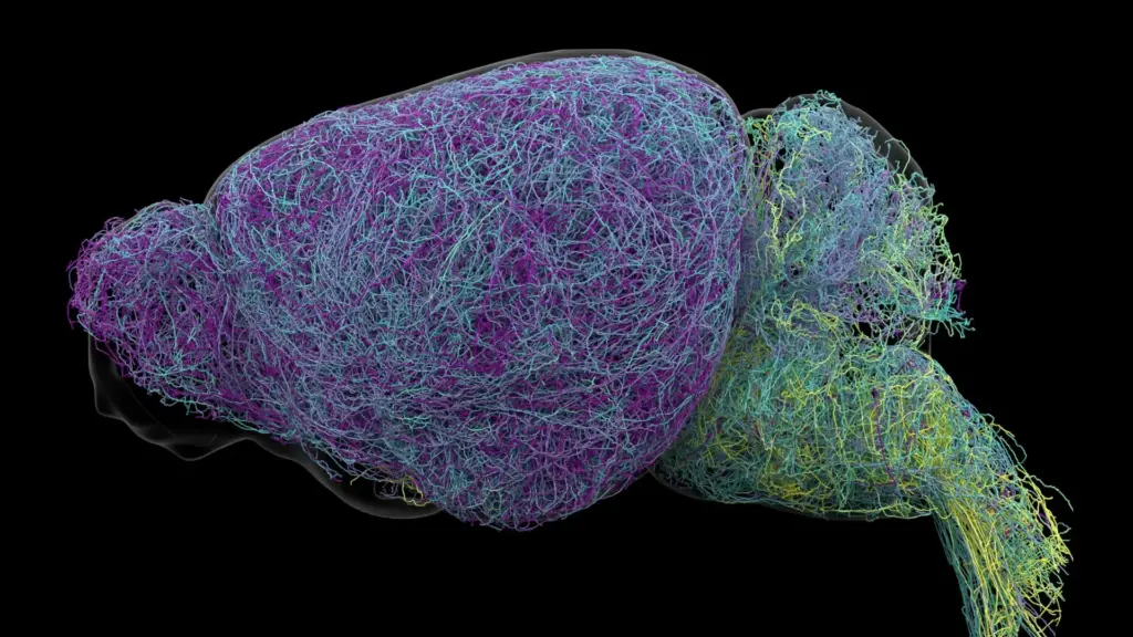
The breakthrough began with an exhaustive effort to map the anatomical architecture of the LC. By tracing the connections of individual neurons, the research team identified that specific groups of cells are hardwired to communicate with distinct regions of the nervous system. This revelation suggests that the LC is not merely flooding the brain with a singular chemical signal, but rather acting as an intricate, targeted postal network.

The spatial organization of these neurons is key to their specialized roles. The study revealed that neurons located in the upper, or dorsal, portion of the locus coeruleus primarily project their axons toward the cerebral cortex and the forebrain. In contrast, cells situated in the lower, or ventral, portion of the LC direct their signals toward the brainstem and the spinal cord. This structural arrangement allows different sub-regions of the LC to exert influence over separate brain functions, effectively allowing the brain to manage complex, multifaceted environments with remarkable precision.
"The findings suggest the brain’s norepinephrine system is far more like a targeted postal network than a foghorn," said Karel Svoboda, director of Neural Dynamics at the Allen Institute and a co-author of the study. By moving away from the "foghorn" model, scientists can now appreciate how the brain manages to prioritize certain information while filtering out irrelevant noise—a function essential for survival and cognitive performance.
Why the Discovery Could Matter for Brain Disorders
The implications of this discovery extend well beyond basic biology. Because norepinephrine is central to the regulation of attention, mood, and stress, it has long been a primary target for pharmacological interventions. Medications used to treat clinical depression, generalized anxiety, and Attention Deficit Hyperactivity Disorder (ADHD) often attempt to modulate norepinephrine levels.
Current clinical approaches, however, are often blunt instruments. Because these treatments typically influence norepinephrine across multiple brain circuits simultaneously, they frequently result in a trade-off: a patient might experience improved focus or mood, but simultaneously suffer from unwanted side effects due to the medication’s impact on unrelated brain regions. By providing a clearer, more granular map of the locus coeruleus’s specialized pathways, this research offers a roadmap for the development of next-generation therapies. If scientists can design drugs that target specific circuits rather than the entire norepinephrine system, they may be able to treat symptoms more effectively while minimizing systemic side effects.
Furthermore, the locus coeruleus is known to be one of the first areas of the brain to show signs of deterioration in patients suffering from Alzheimer’s disease. As researchers work to understand the progression of neurodegenerative conditions, mapping the specific pathways that fail in the early stages of disease could prove critical for early diagnosis and the development of targeted, neuroprotective treatments. This study was supported by the National Institutes of Health’s Brain Research Through Advancing Innovative Neurotechnologies (BRAIN) Initiative, which aims to catalyze the development and application of innovative technologies to produce a revolutionary new dynamic picture of the human brain.
Scientists Identify Brain Cells Involved in Learning
Beyond the structural mapping, the researchers uncovered functional distinctions that further clarify how the LC regulates behavior. Using behavioral experiments in mice, the team observed how different neurons responded to learning tasks. When mice were required to perform decision-making tasks—specifically, when they had to adjust their choices following an unfavorable outcome—the neurons in the dorsal LC showed a marked increase in activity. This confirmed that the dorsal-to-cortex pathway is intimately involved in the cognitive process of learning and adaptive decision-making.
In contrast, neurons in the ventral LC displayed a completely different behavioral signature. Their activity spiked shortly before the mice chose to ignore cues that indicated a potential reward. This suggests that the ventral region acts as a gatekeeper, regulating whether an animal decides to engage with its environment or ignore available opportunities.
"What emerged was a clear map: neurons in the dorsal LC that send signals upward to the cortex are involved in learning, whereas neurons in the ventral region projecting downward to the brainstem and spinal cord govern whether animals engage with their environment at all," explained Svoboda. He further noted that these functional and anatomical differences are reinforced by distinct gene expression patterns, proving that the specialization of these cells is embedded in their very genetic makeup.
Researchers Discover a Record-Breaking Neuron
To achieve this level of detail, the researchers performed an unprecedented examination of the mouse brain. The project involved whole-brain imaging of nearly 35,000 neurons and the analysis of genetic profiles for almost 400,000 individual cells. Perhaps most strikingly, the team successfully reconstructed the full physical architecture of several exceptionally complex nerve cells.
During this process, they encountered a structural anomaly: LC neurons possess axons of extraordinary length. Axons are the long, slender projections that carry electrical impulses away from the cell body to other neurons. While most neurons have relatively short, localized reaches, the researchers found that LC axons measured, on average, approximately 35 centimeters—an immense distance for a structure within a mouse brain. The most impressive specimen reached a reconstructed length of 70.32 centimeters, making it part of the longest single neuron ever measured in a mouse.
Jeremiah Cohen, a scientist at the Allen Institute and co-author of the study, noted the physiological peculiarity of these cells. "This neuron, like many others that we studied, supplies norepinephrine to a very large volume of the cerebral cortex. For the brain, this is highly unusual. Most neurons are more specific in their targets," said Cohen. However, the researchers also observed that despite this massive reach, the neuron remained highly selective. It did not blindly disperse norepinephrine everywhere; rather, it deliberately bypassed the cerebellum, brainstem, and spinal cord. This reinforces the core finding of the study: even at the extreme physical scale of these neurons, the brain’s signaling remains highly organized and selective rather than diffuse.
A New Understanding of How the Brain Learns
Finally, the study identified intriguing parallels between the norepinephrine system and the brain’s dopamine signaling system. Dopamine is widely recognized for its role in the basal ganglia, a region of the brain critical for habit formation. The researchers suspect that these two chemical systems—dopamine and norepinephrine—operate in tandem to facilitate learning. By working together, they may allow the brain to process and acquire multiple complex and abstract concepts simultaneously.
By revealing the intricate, specialized, and highly physical nature of the neurons within the locus coeruleus, this research provides a far more detailed picture of how the mammalian brain communicates and adapts to its environment. While these findings are currently derived from experiments in mice, they provide a foundational framework for understanding human neurobiology. As researchers continue to decode these pathways, the prospect of designing treatments that interact with the brain’s specific, localized circuits—rather than the entire system—moves closer to reality, promising a more nuanced approach to treating the neurological and mental health conditions that affect millions of people worldwide.