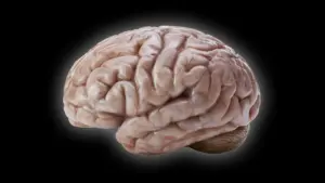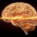








Deep within the brainstem lies a tiny, unassuming cluster of nerve cells that wields an outsized influence over the human experience. This region, known as the locus coeruleus (LC)—a term derived from the Latin for "blue place"—has long been a subject of fascination for neuroscientists. Despite containing only a small fraction of the brain’s total neuronal population, the LC serves as the primary engine for the production of norepinephrine (NE). This vital chemical messenger acts as a biological conductor, orchestrating the body’s complex reactions to stress and excitement while simultaneously regulating fundamental processes such as heart rate, learning, and the capacity for sustained attention.
For decades, the prevailing scientific consensus characterized the locus coeruleus as a "loudspeaker" for the brain. Researchers assumed that the region functioned as a broadcast system, sending a uniform, undifferentiated norepinephrine signal across the entire brain to keep the nervous system in a state of general arousal. However, a groundbreaking study recently published in the journal Nature has fundamentally challenged this long-held understanding. By utilizing a sophisticated combination of advanced brain imaging, high-resolution neural recordings, cutting-edge genetic techniques, and rigorous behavioral experiments in mice, researchers at the Allen Institute have revealed that the LC is far more nuanced than previously imagined. Rather than acting as a singular broadcaster of generic information, the LC communicates through a series of highly distinct, specialized pathways, with different groups of neurons firing in response to specific experiences and stimuli.
A Hidden Brain Network That Sends Precise Messages
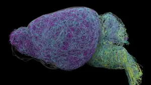
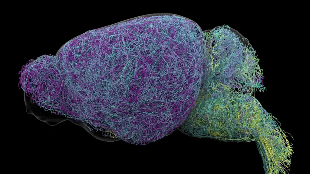
The discovery highlights a sophisticated level of organization that was previously unrecognized by the scientific community. By painstakingly tracing the anatomical connections of individual LC neurons, the researchers discovered that the region acts less like a megaphone and more like a targeted, highly efficient postal network. Different cohorts of cells are dedicated to communicating with specific regions of the nervous system, ensuring that norepinephrine is released only where it is needed, rather than being flooded throughout the entire brain.

This structural organization is surprisingly consistent. Neurons located in the upper, or dorsal, portion of the locus coeruleus are primarily wired to connect with the cerebral cortex and the forebrain—areas of the brain associated with higher-order thinking and cognition. In contrast, cells situated in the lower, or ventral, portion of the region send their projections downward, primarily targeting the brainstem and the spinal cord. This clear physical separation allows the different segments of the LC to exert influence over entirely different physiological and psychological functions.
"The findings suggest the brain’s norepinephrine system is far more like a targeted postal network than a foghorn," noted Karel Svoboda, director of Neural Dynamics at the Allen Institute and a co-author of the study. By identifying these specific circuits, the research team has mapped a level of neurological precision that suggests the brain is capable of micro-managing its chemical signaling in ways that were previously thought impossible.
Why the Discovery Could Matter for Brain Disorders
The implications of this research extend far beyond basic anatomical mapping. Understanding the precise architecture of the norepinephrine signaling network could eventually revolutionize the way clinicians approach a wide array of neurological and mental health conditions. Because norepinephrine plays a central role in modulating mood and attention, it is already a critical target for many common medications used to treat conditions such as depression, anxiety, and Attention Deficit Hyperactivity Disorder (ADHD).
Furthermore, the locus coeruleus is one of the very first regions of the brain to exhibit signs of deterioration in patients suffering from Alzheimer’s disease. The fact that these cells are so early to falter suggests that the new insights into their organization could provide a roadmap for understanding the progression of neurodegenerative diseases. Currently, many pharmacological treatments that target the norepinephrine system suffer from a lack of selectivity; they tend to affect multiple brain circuits simultaneously. This "blunt instrument" approach often results in a trade-off where a patient might experience relief from one set of symptoms while enduring unwanted side effects elsewhere. With a more detailed map of the LC’s specialized pathways, scientists hope to eventually design more precise therapies—drugs that can influence specific circuits involved in particular symptoms without disrupting the entire signaling network. This research was supported by the National Institutes of Health’s Brain Research Through Advancing Innovative Neurotechnologies (BRAIN) Initiative, a project dedicated to developing new tools to revolutionize our understanding of the human brain.
Scientists Identify Brain Cells Involved in Learning
Beyond the structural mapping, the researchers uncovered functional distinctions that further clarify how the LC contributes to cognitive processing. The study revealed a clear divergence in the roles played by dorsal and ventral neurons. Cells in the dorsal LC, which project to the cortex, showed a marked increase in activity during learning tasks. When the mice were required to engage in decision-making and, crucially, when they had to adjust their choices following an unfavorable or unexpected result, these neurons became highly active.
In contrast, the ventral LC neurons exhibited a completely different behavioral pattern. Their activity increased shortly before the mice chose to ignore external signals—such as cues indicating that a reward might be available. This suggests that the ventral portion of the LC plays a role in gating behavior, effectively regulating whether an animal chooses to engage with its environment or ignore potential opportunities.
"What emerged was a clear map: neurons in the dorsal LC that send signals upward to the cortex are involved in learning, whereas neurons in the ventral region projecting downward to the brainstem and spinal cord govern whether animals engage with their environment at all," Svoboda explained. The researchers also confirmed that these functional differences are not merely the result of location; they are supported by distinct gene expression patterns, indicating that the two groups of cells are biologically specialized at the molecular level.
Researchers Discover a Record-Breaking Neuron
The scale of the study was immense, involving an exhaustive examination of the mouse brain. The team utilized whole-brain imaging to analyze nearly 35,000 individual neurons and performed detailed genetic profiling on nearly 400,000 cells. Perhaps most impressively, they successfully reconstructed the complete, three-dimensional structures of several exceptionally complex nerve cells.
During this reconstruction, the team encountered a remarkable finding: LC neurons possess axons of extraordinary length. Axons are the thin, thread-like projections that allow nerve cells to transmit electrical impulses to other parts of the nervous system. The researchers found that, on average, LC axons measured approximately 35 centimeters in length—an immense distance for a mouse. Even more astounding was the discovery of a single neuron with a reconstructed length of 70.32 centimeters, making it among the longest single neurons ever measured in a mouse.
"This neuron, like many others that we studied, supplies norepinephrine to a very large volume of the cerebral cortex," said Jeremiah Cohen, a scientist at the Allen Institute and co-author of the study. "For the brain, this is highly unusual. Most neurons are more specific in their targets." However, Cohen noted that even this record-breaking cell maintains a degree of selectivity. Despite its vast reach across the cortex, the neuron does not broadcast its signal indiscriminately; it notably ignores the cerebellum, brainstem, and spinal cord. This observation further reinforces the core theme of the study: the locus coeruleus is a highly organized, selective system rather than a disorganized source of general background noise.
A New Understanding of How the Brain Learns
Finally, the study identified intriguing parallels between the norepinephrine system and the brain’s dopamine signaling pathways. Dopamine is widely known for its role in the basal ganglia, a region of the brain critical for habit formation and reward-seeking. Scientists now suspect that these two chemical systems—norepinephrine and dopamine—work in tandem to support the learning process. This synergistic relationship may provide the biological infrastructure that allows the brain to acquire, process, and integrate multiple complex or abstract concepts simultaneously.
By mapping the intricate connections, specialized functional roles, and the surprising physical dimensions of the neurons within the locus coeruleus, the research team has provided a much higher-resolution picture of how the brain communicates and learns. While these findings are derived from experiments in mice, they provide a foundational framework that could fundamentally alter the study of human neurobiology. By shifting away from the idea of the LC as a uniform "foghorn" and embracing the reality of a precise, multi-pathway network, the medical community may one day be better equipped to design targeted interventions for the most challenging brain disorders of our time.