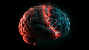
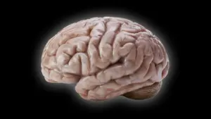








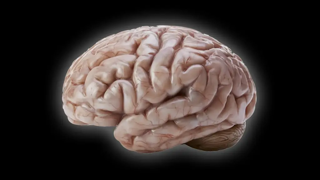
Most primates are characterized by their remarkably complex brains, often dominated by intricate folds and grooves known as gyri and sulci. These structures are more than just anatomical features; they are the architectural hallmark of advanced cognitive function, providing a massive surface area that houses billions of neurons. Yet, nature has crafted significant exceptions to this rule. Among them is the common marmoset (Callithrix jacchus), a small South American monkey whose brain remains surprisingly smooth throughout its life.
For years, the stark contrast between the walnut-like, folded human brain and the smooth, streamlined brain of the marmoset has captivated evolutionary biologists. Now, a team of researchers at the German Primate Center (DPZ)—the Leibniz Institute for Primate Research in Göttingen—has unlocked key cellular mechanisms that explain this divergence. Their findings, published in the journal Science Advances, offer a window into the evolutionary history of primates and provide a new framework for understanding the biological foundations of human brain development.
Why Some Primate Brains Are Smooth Instead of Folded
Brain morphology is one of the most critical factors distinguishing primate species. In humans, the cerebral cortex is highly convoluted, a design that maximizes surface area within the constrained space of the cranium. This increased surface area is essential for housing the vast networks of neurons that facilitate high-level reasoning, complex social behavior, and advanced sensory processing.

Evolutionary scientists have long maintained that the common ancestor of modern primates likely possessed a moderately sized brain that already exhibited some degree of folding. However, as evolutionary pressures shaped different lineages, some species veered away from this path. The common marmoset, which has become a vital model in modern biomedical research, presents a unique case study in brain evolution. Unlike their larger-brained cousins, these monkeys exhibit a nearly lissencephalic—or smooth—cerebral cortex.
To decode the mystery of why these brains develop so differently, researchers at the German Primate Center conducted a rigorous comparative analysis. "We wanted to understand which changes at the cellular level cause the marmoset brain to grow less and form fewer folds," says Lidiia Tynianskaia, a PhD student in the Junior Research Group Brain Development and Evolution at the DPZ and co-first author of the study.
The team observed that at the very onset of embryogenesis, the marmoset brain actually follows a trajectory that mimics that of larger, folded primate brains. The fundamental composition and initial structure are remarkably similar. "As development progresses," Tynianskaia explains, "processes must therefore occur that effectively slow down the production of nerve cells." This shift, they hypothesized, is the key to the marmoset’s eventual brain size and surface morphology.
Scientists Discover What Slows Brain Growth
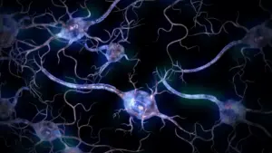
The research team turned to an innovative methodology to test their hypothesis: brain organoids. These three-dimensional, miniature brain-like structures are grown from stem cells and serve as sophisticated models that replicate the fundamental features of early human and marmoset brain development. By comparing these organoids, the researchers were able to zoom in on the neural progenitor cells—the "factory" cells responsible for churning out neurons.
The size and complexity of the cerebral cortex are inextricably linked to how these progenitor cells function. If these cells are highly prolific and divide rapidly over an extended period, they produce a massive population of neurons, which in turn necessitates the expansion and folding of the cortex. Conversely, if these cells are constrained in their proliferation or their developmental timing is truncated, the resulting brain will be smaller and smoother.
The DPZ team discovered that the marmoset’s smaller, smoother brain is not the consequence of a single genetic "switch," but rather the result of a multi-faceted cellular slowdown. "Our investigations have shown that certain progenitor cells in the common marmoset divide significantly more slowly than in humans," notes César Mateo Bastidas Betancourt, a PhD student and co-first author of the study.
The researchers identified two primary issues within these progenitor cell populations. First, the cells themselves exhibit structural differences; they possess fewer processes compared to their human counterparts, which limits their proliferative capacity. Second, the fundamental rate of division is sluggish, meaning that for every hour of development, fewer new neurons are added to the growing brain.
Beyond the rate of division, the temporal aspect of development also proved critical. The team observed that the "window of opportunity"—the timeframe during which these progenitor cells can rapidly multiply—is significantly shorter in marmosets than it is in humans. When these three factors—slow division rates, simpler cell architecture, and a truncated growth window—are combined, the result is a massive reduction in total neuron production, leading directly to the lack of cortical folding seen in the adult marmoset.
Miniature Brains Reveal Important Developmental Clues
The use of organoids was pivotal in reaching these conclusions. While traditional animal models have their place in neuroscience, organoids allow researchers to perform large-scale, statistically robust experiments that would be impossible to conduct in live animal subjects. However, the team was careful to validate their findings against reality. To ensure that the organoid results were not mere artifacts of the laboratory, they cross-referenced their findings with fetal brain tissue samples.
One of the most significant hurdles in using organoids is determining whether they grow at the same speed as an actual brain. "The study combines the advantages of in vivo and in vitro methods," explains Michael Heide, head of the Junior Research Group Brain Development and Evolution. "Organoids are well-suited for obtaining statistically robust results because such sample sizes are not feasible in primates. We subsequently repeated some key experiments in fetal brain tissue to confirm the results from the organoids."
The team successfully established a crucial developmental timeline, finding that a marmoset brain organoid grown for 50 days closely matches the developmental stage of a natural marmoset brain at 90 days. This correlation was essential for the researchers to interpret their findings with confidence. By establishing this chronological map, they could accurately compare the behavior of cells in the dish with the biological realities of primate gestation.
What Smooth Monkey Brains Reveal About Human Brain Evolution
The implications of this study extend far beyond the anatomy of the marmoset. By identifying the precise cellular levers that dictate brain size and folding, the researchers have provided a clearer picture of the evolutionary mechanisms that have shaped the primate order. It appears that the vast diversity of primate brain architecture—ranging from the smooth brains of small monkeys to the highly convoluted brains of great apes and humans—is driven by subtle, finely tuned adjustments to the behavior and timing of progenitor cells.
These discoveries are particularly valuable for the broader field of neuroscience. Understanding the mechanisms that govern neuron production is fundamental to understanding human health. When these developmental processes go awry in humans, the result can be severe neurodevelopmental disorders, such as microcephaly or cortical malformations.
By pinpointing how the marmoset brain limits its own growth, scientists are gaining a better understanding of the biological "brakes" and "accelerators" that regulate brain size. This knowledge serves as a foundational step toward understanding the human condition. As researchers continue to explore the differences between species, the "smooth" brain of the marmoset serves as a vital mirror, helping scientists identify the unique cellular processes that allowed the human brain to expand, fold, and ultimately support the complex cognitive abilities that define our species. Through these miniature, laboratory-grown models, the path toward better diagnostics and potential future interventions for developmental disorders becomes increasingly clear.