







For decades, the medical community has approached clinical depression as a monolithic diagnosis. Patients experiencing a persistent low mood, overwhelming anxiety, or a loss of interest in life are frequently funneled into the same diagnostic category: Major Depressive Disorder (MDD). Yet, clinicians have long observed that this "one-size-fits-all" label masks a deeply heterogeneous reality. One patient may struggle primarily with debilitating, repetitive negative thoughts, while another might battle substance abuse or the lingering shadows of past trauma.
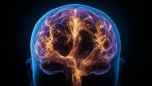
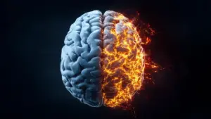
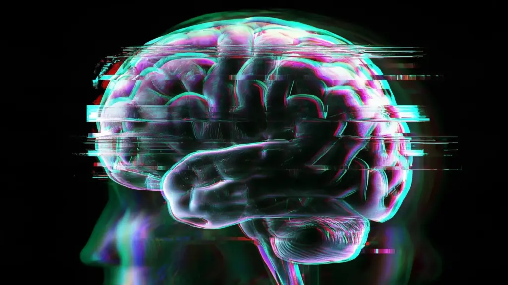
Now, a pioneering study conducted by researchers at the University of Helsinki has provided compelling biological evidence to explain this clinical diversity. By examining the brain activity of 263 patients diagnosed with depression, scientists have uncovered that the condition is not characterized by a single, consistent brain signature. Instead, depression may manifest through at least five distinct patterns of brain function, with some patients displaying hyper-connected neural networks while others exhibit marked reductions in communication between brain regions.
A Global Health Challenge
The implications of these findings are significant, particularly given the scale of the crisis. Depression remains a major global health concern, touching the lives of millions. According to World Health Organization data from 2025, approximately 332 million adults—roughly 5.2% of the global adult population—are currently living with the condition. The burden is particularly heavy in Finland, where depression stands as the leading individual cause for both extended sick leave and long-term disability pensions.
Despite the prevalence of the disorder, current treatment protocols often rely on a trial-and-error approach. Physicians prescribe antidepressants or recommend specific psychotherapies, but because patients respond so differently, finding the right path to recovery can be a lengthy, frustrating process. The Helsinki study offers a glimpse into a future where these decisions might be informed by objective, biological data.
Decoding the Biology of Mood
To investigate the biological underpinnings of depression, the research team, led by Director Satu Palva of the Neuroscience Center at the University of Helsinki, compared 263 patients with MDD against a control group of 75 healthy participants. The primary focus was on "functional connectivity"—a measure of how closely the electrical activity in different brain regions is coordinated. When brain regions show synchronized activity, it serves as a sign that they are working together as part of a larger, integrated network.
The results were striking. The team identified five distinct groups of patients based on their specific connectivity patterns. As Director Palva explains, the contrast was particularly noteworthy because it occurred within the same diagnostic category. In some individuals, the communication between brain regions was abnormally strong, while in others, it was significantly weaker than what was observed in the healthy control group.
This discovery may finally resolve a long-standing mystery in neuroscience: why previous studies have so often reached conflicting conclusions. If different researchers inadvertently study disparate groups of patients—each possessing a different biological "profile" of depression—it is inevitable that they will observe different patterns of brain activity. This study suggests that the brain of a person with depression is not "broken" in one uniform way; rather, it is altered in ways that depend entirely on the specific nature of their depression.
Five Profiles of Brain Function
The researchers categorized the participants into five distinct groups, each defined by characteristic patterns of connectivity and correlated with specific clinical symptoms.
The first group exhibited relatively strong connections between brain regions. This profile was linked to a more severe symptom burden, including heightened anxiety, persistent rumination—the act of repeatedly dwelling on negative thoughts or past distress—and a reduced capacity to function in daily life.
The second group presented a starkly different picture: relatively weak communication between brain regions. Interestingly, participants in this group generally experienced the mildest symptoms of the cohort. This stands in contrast to the third group, which also showed widespread weak connectivity but was specifically defined by the prominence of post-traumatic stress disorder (PTSD) symptoms.
The fourth group showcased a more complex, "mixed" profile. These patients demonstrated a combination of unusually strong connections in certain brain regions alongside weaker connections in others. This group was associated with more severe depressive symptoms, lower overall well-being, and greater challenges with substance abuse.
Finally, the fifth group represented the extreme end of the spectrum regarding connectivity. Participants in this group had the strongest connections between brain regions of any group identified in the study. Substance abuse was a particularly prominent feature for these individuals, while their trauma-related symptoms were less pronounced compared to the other groups.
These findings suggest that the differences go far beyond simple "strong" or "weak" connectivity. The groups also differed in which specific brain regions were involved and the frequencies at which their activity was synchronized, indicating that the neural underpinnings of depression are multifaceted and deeply varied.
Precision Through Magnetoencephalography
The precision of these findings is largely attributed to the use of magnetoencephalography (MEG). Unlike traditional functional magnetic resonance imaging (fMRI), which tracks slower changes in blood flow, MEG detects the extremely faint magnetic fields produced by the brain’s electrical activity. Because it can record these signals with millisecond precision, the research team was able to follow the rapid timing and coordination of communication between brain regions as it happens in real-time.
"MEG enabled us to monitor electrical brain activity with millisecond precision, helping us get closer to what actually happens in the brain at any given moment," says Palva. "Previously, depression phenotypes have been studied using methods with slower responses." By capturing these fast signals, the team could identify subtle, rapid changes in brain activity that would be invisible to slower measurement techniques.
The Path Toward Personalized Psychiatry
While the identification of these five profiles is a major step forward, the researchers are careful to manage expectations regarding immediate clinical applications. We are not yet at the stage where a quick brain scan can dictate a specific antidepressant prescription or a tailored therapy regimen. However, the study provides a clear, evidence-based roadmap for the future of mental health care.
The long-term goal is to bridge the gap between biological patterns and the lived experiences of patients. By linking these distinct brain signatures to specific clusters of symptoms, researchers hope to move away from the "trial-and-error" model of psychiatric care. If a clinician can identify a patient’s specific brain profile, they might eventually be able to select treatments that are statistically more likely to succeed for that individual, thereby reducing the time spent struggling with ineffective interventions.
For now, the study stands as a powerful reminder that depression is not a single, monolithic disease. It is a diverse condition involving markedly different biological changes, sometimes occurring in entirely opposite directions within the brain. By shifting the focus toward these distinct biological signatures, the researchers have opened a new window into the complexity of the human mind, offering hope that more precise, personalized, and effective care is on the horizon.
This research, conducted in collaboration with Aalto University and the Helsinki and Uusimaa Hospital District (HUS), underscores the necessity of interdisciplinary efforts to tackle one of the most pervasive health challenges of our time.