








Two individuals walking into a clinician’s office with a diagnosis of major depressive disorder may share the same clinical label, but their lived experiences can be worlds apart. One patient might grapple with the relentless cycle of repetitive negative thoughts and debilitating anxiety, while another may find themselves struggling with the long-term echoes of trauma or the destructive pull of substance abuse. For decades, the psychiatric community has operated under the assumption that these diverse manifestations of depression might share a common underlying biological signature. However, groundbreaking new research from the University of Helsinki suggests that the reality is far more complex, revealing that depression is not a singular biological entity but a multifaceted condition characterized by vastly different patterns of brain activity.
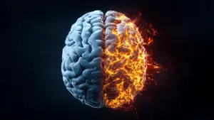
The study, which investigated the neural architecture of depression using advanced imaging technology, has uncovered a striking reality: people diagnosed with the same clinical condition exhibit dramatically different, and sometimes even opposite, patterns of brain function. In some individuals, the communication between critical brain regions was found to be unusually hyperactive, while in others, that same communication was significantly dampened. These findings provide a compelling new perspective on why depression manifests in such varied ways and why treatment responses can be so unpredictable.
The Global Scope of a Complex Challenge
Depression remains one of the most pressing health challenges of the modern era. According to data from the World Health Organization (2025), approximately 332 million adults globally—roughly 5.2% of the population—are currently living with the condition. The impact is profound, affecting not just the individual’s emotional well-being but their ability to function in daily life, maintain employment, and foster relationships.

In Finland, the socio-economic impact is particularly stark, with depression standing as the single leading cause of both extended sick leave and disability pensions. Given these high stakes, the pressure to understand the biological mechanisms driving the disorder has never been greater. By moving beyond the symptom-based diagnosis, researchers hope to shift the paradigm toward a more nuanced, biologically informed approach to mental health care.
Deciphering the Biological Landscape of Depression
To unravel the biological mysteries hidden within the depressed brain, researchers at the University of Helsinki embarked on an ambitious study involving 263 participants diagnosed with major depressive disorder and a control group of 75 healthy volunteers. The primary goal was to measure functional connectivity—a metric that tracks how closely activity in various brain regions is synchronized. When regions fire in unison, it indicates they are functioning as a cohesive network. However, the study serves as a critical reminder that "stronger" connectivity is not inherently "better." The brain is a delicate balance of activation and inhibition, and deviations in either direction can lead to profound dysfunction.
Director Satu Palva of the Neuroscience Center at the University of Helsinki, who led the research, noted that the findings challenge long-held assumptions about the uniformity of the disorder. "What was particularly interesting was the contrasting patterns of brain activity found under the umbrella of the same depression diagnoses," Palva explains. "In some individuals, the functional connectivity between brain regions was stronger than usual, while in others it was weaker."
This discovery offers a potential explanation for why previous neurological studies on depression have often yielded conflicting or inconsistent results. If researchers are inadvertently grouping patients with fundamentally different neurological profiles into a single study, the resulting data is bound to be noisy and contradictory. These findings suggest that the clinical diagnosis of depression is, in many ways, an umbrella term that covers several distinct biological subtypes.
Five Distinct Profiles of Brain Connectivity
The researchers successfully classified the participants into five distinct groups, each defined by a specific signature of brain connectivity and a corresponding set of clinical symptoms. This classification system provides a much-needed map for understanding how biological differences translate into human behavior.
The first group exhibited relatively strong connectivity across various brain networks. This profile was closely tied to more severe clinical presentations, including intense anxiety, constant rumination—the repetitive, obsessive dwelling on negative thoughts or past distress—and a significantly reduced capacity to manage the demands of daily life.
In contrast, the second group presented with generally weaker communication between brain regions. Perhaps surprisingly, this group reported the mildest symptom profiles compared to their peers in the study, suggesting that reduced connectivity does not necessarily correlate with the most severe clinical outcomes.
The third group demonstrated a more widespread pattern of weak connectivity across significant portions of the brain. This cluster was particularly noteworthy for the prominence of post-traumatic stress disorder (PTSD) symptoms, suggesting that the neurological signature of trauma-related depression may be fundamentally different from other forms of the disorder.
The fourth group displayed a "mixed" connectivity profile, characterized by a chaotic combination of hyperactive and hypoactive regions. These participants typically experienced a complex array of challenges, including severe depression, high levels of substance abuse, and a general decline in overall well-being.
Finally, the fifth group showed the most intense connectivity of any cohort in the study. Substance abuse was a defining feature for these individuals, while their trauma-related symptoms were noticeably less pronounced than those found in the third group.
Importantly, every one of these five groups showed clear and distinct differences from the healthy control participants. The deviations were not limited to the strength of the connections; they also varied in which specific brain regions were involved and the frequency at which those regions synchronized. This suggests that the internal "rhythm" of the brain is fundamentally altered in depression, and these alterations manifest in highly individualistic ways.
Measuring the Millisecond Pulse of the Brain
The ability to identify these nuances was made possible by the use of magnetoencephalography (MEG). While traditional imaging methods like functional MRI are excellent at showing which parts of the brain are active, they are relatively slow, capturing changes over several seconds. In contrast, MEG detects the incredibly faint magnetic fields produced by the brain’s electrical activity, allowing researchers to track signals with millisecond precision.
"MEG enabled us to monitor electrical brain activity with millisecond precision, helping us get closer to what actually happens in the brain at any given moment," Palva notes. "Previously, depression phenotypes have been studied using methods with slower responses." This high temporal resolution was essential for identifying the rapid, flickering changes in connectivity that define these five distinct neural profiles. By capturing the brain in motion, the researchers were able to see not just where the brain was working, but how its various regions were coordinating their fast-paced communication.
A New Horizon for Personalized Psychiatry
The potential implications of these findings for the future of mental health treatment are profound. Currently, the process of treating depression is often a matter of trial and error; a patient might try several different medications or therapy modalities before finding one that provides genuine relief. Because the underlying biology varies so wildly between individuals, what works for one person may be entirely ineffective for another.
While the researchers are careful to emphasize that this technology is not yet ready for the clinic—a patient cannot currently walk into a doctor’s office and request a brain scan to determine their prescription—the study maps a clear path forward. The ultimate goal is to refine these biological markers to the point where they can assist clinicians in selecting the most effective treatment from the outset. By matching a patient’s specific neural profile to the therapy most likely to address their particular brain dysfunction, clinicians could significantly reduce the time and suffering involved in finding an effective treatment.
For now, the study stands as a significant contribution to our understanding of the human brain. It challenges the medical community to look past the label of "depression" and recognize that the condition is a heterogeneous spectrum of neurological states. By acknowledging that these biological differences exist, the field moves one step closer to a future where psychiatric care is as precise and personalized as oncology or cardiology. The research was carried out through a collaborative effort between the University of Helsinki, Aalto University, and the Helsinki and Uusimaa Hospital District (HUS), marking a collaborative milestone in the ongoing quest to decode the complexities of the human mind.