




Why do some new skills seem to click almost instantly while others remain frustratingly difficult even after repeated practice? For decades, the answer has been attributed primarily to individual talent, the intensity of focus, or the sheer number of hours spent in deliberate practice. However, a groundbreaking study suggests the secret to mastery may lie in a physiological "readiness" of the brain—a state that can be externally influenced by the body’s internal signaling systems.
New research from Tohoku University, published on August 25, 2026, in the journal iScience, reveals that stimulating the vagus nerve—a major communication highway between the body and the brain—can significantly strengthen long-term motor learning. By demonstrating that this nerve stimulation can act as a catalyst for memory consolidation, researchers have uncovered a previously overlooked dimension of how our physical systems help new skills persist long after the training session ends.
The Vagus Nerve as a Learning Accelerator
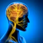
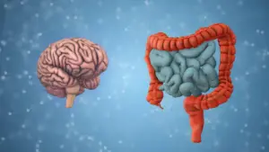
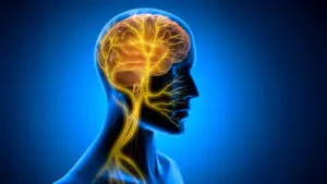
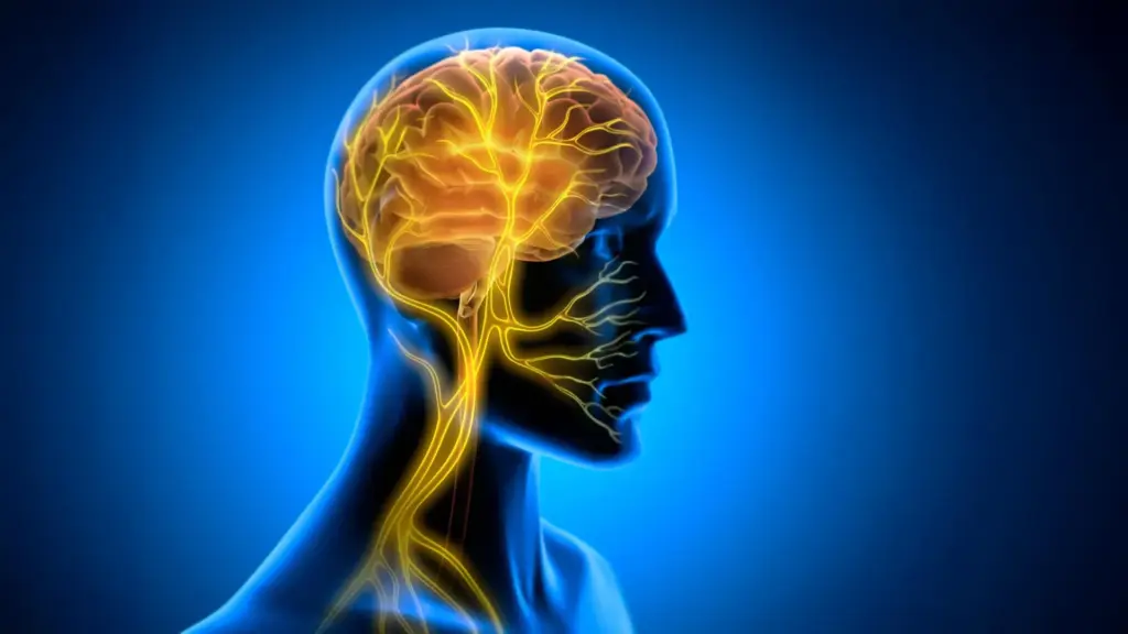
The vagus nerve is one of the most critical components of the autonomic nervous system, serving as a bidirectional conduit for information. It constantly transmits sensory data from the internal organs to the brain, while simultaneously carrying regulatory instructions from the brain back to those organs. Because of its expansive reach and its role in modulating physiological states, the vagus nerve has long been a target for medical intervention.

Vagus Nerve Stimulation (VNS) is already a clinically approved therapy for several neurological and psychiatric conditions, including epilepsy and treatment-resistant depression. Traditionally, scientists have understood the mechanism of VNS through the lens of neuromodulation, focusing on how electrical pulses alter the activity of neurotransmitter systems, such as norepinephrine and acetylcholine, which are essential for focus and plasticity.
However, the team at Tohoku University, which specializes in the super-network physiology of the brain, suspected there was more to the story. Their findings suggest that VNS does not merely tweak chemical levels; it may fundamentally change the vascular environment of the brain, creating a rhythmic, metabolic pulse that prepares neural circuits for lasting change.
The "Post-Practice" Window of Opportunity
To test their hypothesis, the researchers conducted a series of experiments using mice. They developed a specialized, miniaturized cuff electrode capable of being permanently attached to the left cervical vagus nerve. With this device, they could precisely control the timing and intensity of stimulation during a controlled learning task.
The researchers focused on a task known as horizontal optokinetic response (HOKR) learning. This is a cerebellum-dependent skill that requires mice to track moving visual stripes, effectively improving their ability to coordinate eye movements. This task serves as a reliable model for human motor learning, mirroring the automatic, subconscious adjustments our eyes make when we stand on a train platform and watch the world move past us.
Crucially, the study design separated the stimulation from the training itself. The researchers applied VNS only after each training session, rather than during the performance of the task. This distinction proved to be the pivotal insight of the study.
The stimulation did not yield any immediate performance boost during the training sessions. The mice did not appear "smarter" or more adept while the stripes were moving. However, the effects became strikingly apparent on subsequent days. Mice that received the post-training VNS demonstrated significantly stronger, more durable long-term learning compared to the control group.
"The key point is that VNS was delivered only after training," explains Professor Ko Matsui of Tohoku University. "Our findings suggest that VNS may open a hidden window of opportunity for enhanced learning by making the brain environment more receptive to long-lasting change."
This implies that the brain requires a specific physiological state to consolidate short-term practice into long-term memory. By stimulating the vagus nerve after the work is done, the researchers were essentially "tagging" the experience for storage, ensuring that the neural pathways engaged during practice were locked in more firmly.
Rhythmic Vascular Changes: A New Mechanism
To understand exactly how the vagus nerve facilitates this consolidation, the research team investigated the physiological changes occurring within the brain after stimulation. They utilized fiber photometry to measure blood volume activity near the cerebellar flocculus—the specific region of the cerebellum responsible for HOKR learning.
The data revealed a complex, two-phase vascular response. Following a single round of VNS, local blood volume in the cerebellar region initially decreased before experiencing a delayed, sustained increase. When the researchers applied repeated stimulation, they observed the creation of rhythmic oscillations in blood volume.
The team found a compelling correlation between these vascular rhythms and the speed of learning. Mice that exhibited larger, more pronounced blood volume oscillations during the post-training period generally showed superior performance by the fifth day of the experiment. This suggests that the "vascular environment"—the rhythmic ebb and flow of blood and, by extension, the metabolic resources delivered to the brain—is a critical, yet previously underestimated, component of the learning process.
"Our brains may be more strongly influenced by the body than we imagine," says Junyu Chen, the lead author of the study. "By tuning the brain’s metabolic environment, including rhythmic vascular movements, we may eventually unlock capacities that would otherwise remain latent."
Implications for the Future of Learning
The discovery that the body plays an active role in "setting the stage" for memory consolidation opens a new frontier in neuroscience. If the brain’s ability to learn is tied to the rhythmic signals sent by the body, then learning is not merely a cerebral activity confined to the skull; it is a whole-body process.
This insight into the brain-body connection has profound implications. If scientists can refine these stimulation protocols, they might be able to create therapeutic interventions for individuals struggling with learning disabilities or those undergoing physical rehabilitation after injuries. By identifying the exact timing and nature of the signals that trigger these vascular rhythms, researchers hope to move toward a future where we can manually "prime" the brain to be more receptive to new information and motor skills.
However, the team acknowledges that this is only the beginning. Future research must focus on the precise protocols required to trigger these benefits in different types of learning and in more complex environments. The researchers aim to map out exactly how this communication between the brain and body supports the architecture of long-term plasticity.
As the scientific community continues to peel back the layers of how we acquire and retain skills, the Tohoku University study serves as a reminder of the body’s hidden influence. Learning may not just be a matter of "practice makes perfect"—it may be a matter of ensuring the body is communicating the right signals to the brain at the right time, effectively turning the practice into permanent, biological change. By better understanding these two-way pathways, researchers hope to not only comprehend how we learn but to eventually enhance the very mechanisms that allow us to master the world around us.