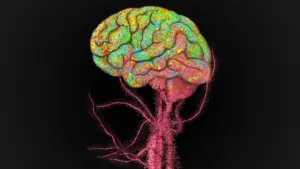








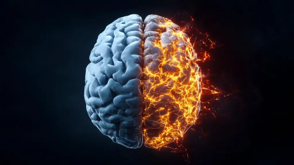
In a significant advancement for neurobiology, researchers at the University of Birmingham have unveiled a promising new strategy to curb harmful brain inflammation by targeting a specific receptor already susceptible to existing pharmacological interventions. This breakthrough, recently published in the journal Brain, centers on the P2X7 receptor, which the study identifies as a primary engine driving neuroinflammatory processes. By successfully blocking this receptor in human brain tissue, the research team has opened a potential pathway toward repurposing established medications to treat a vast array of debilitating neurological and psychiatric conditions.
Led by Professor Nicholas Barnes from the University of Birmingham’s College of Medicine and Health, the research represents a major step forward in the quest to address the inflammation that underlies some of the most persistent challenges in modern medicine. The findings suggest that by modulating the P2X7 receptor, clinicians may eventually be able to reduce the collateral damage associated with conditions ranging from Alzheimer’s disease and Parkinson’s disease to traumatic brain injury (TBI), depression, and psychosis.
Blocking a Key Driver of Brain Inflammation
The focus of the study was the P2X7 receptor, a protein known to play a complex role in the brain’s inflammatory signaling pathways. When the brain experiences injury or stress, inflammatory signaling is a natural, protective response. However, when this process becomes chronic or dysregulated, it can lead to significant neuronal damage and contribute to the progression of neurodegenerative diseases.
To understand the mechanism at play, the researchers employed a two-pronged approach. They utilized live cultures of human brain cells alongside delicate slices of brain tissue harvested during necessary neurosurgical procedures. This allowed the team to observe the interaction between the P2X7 receptor and the inflammatory machinery of the brain in a high-fidelity environment.
The research demonstrated that P2X7 receptors are instrumental in promoting the release of cytokines—the signaling proteins that act as the chemical "messengers" of the immune system. When the cytokines are released in excess, they trigger an inflammatory response that can damage healthy tissue. By introducing a specific antagonist—a chemical compound designed to block the receptor—the researchers observed a profound reduction in the inflammatory response within the human brain tissue.
Professor Nicholas Barnes emphasized the significance of these findings, noting the potential for rapid clinical application. "This exciting discovery marks a major step toward repurposing existing therapeutics to combat neuroinflammation at its source," Barnes stated. "The identification of this receptor could have far-reaching implications for some of the most debilitating and widespread brain disorders such as Alzheimer’s Disease, Parkinson’s and Multiple Sclerosis, or inflammation-linked psychiatric conditions like schizophrenia and depression."
Studying the Brain’s Immune Cells
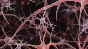
Central to this investigation were microglia, the specialized immune cells of the central nervous system. Microglia are tasked with patrolling the brain, identifying threats, and coordinating the tissue’s response to injury or infection. Under normal conditions, they are essential for maintaining brain homeostasis. However, in the context of disease or trauma, these cells can become overactive, contributing to a "vicious cycle" of inflammation that worsens the initial injury.
A significant hurdle in the field of neuroscience has been the difficulty of studying human microglia in a laboratory setting. Once these cells are removed from the complex, nutrient-rich, and chemically balanced environment of the human brain, they typically lose their defining characteristics and cease to function as they would in vivo. This loss of phenotype has historically made it difficult for scientists to draw reliable conclusions about how human immune cells respond to inflammatory triggers.
To circumvent this, the University of Birmingham team developed a sophisticated method for converting human peripheral monocytes—a type of white blood cell—into microglia-like cells. This innovative technique mirrors a cellular transformation process that has recently been identified as occurring naturally within the human brain during the aging process. By starting with monocytes collected from standard blood samples, the researchers were able to generate a scalable, reliable platform that mimics the behavior of human microglia with unprecedented precision.
This "microglia-in-a-dish" model allowed the researchers to observe how these cells reacted when they were exposed to inflammatory signals and, crucially, how they reacted when that signal was intercepted. When the P2X7 receptor antagonist was applied, the researchers were able to successfully interfere with the signals released by the microglia as the cells entered a state of damage and decay.
"Studying human microglia has long been a major challenge," Professor Barnes explained. "Once removed from their native brain environment, they rapidly lose their defining characteristics, likely due to the absence of critical regulatory signals. Our approach involved the use of monocyte-derived microglia which provide a powerful, scalable, and virtually unlimited platform for studying human microglial biology with unprecedented precision."
From Lab Models to Human Brain Tissue
Having established a functional, high-precision model using the monocyte-derived cells, the team moved to validate their findings in actual human brain tissue. The ability to transition from a laboratory-grown model to living human tissue is a vital step in translational medicine, providing the necessary evidence that an intervention might be effective in a clinical setting.
The researchers tested the application of the P2X7 receptor antagonist on human brain samples obtained through neurosurgical procedures. The results mirrored the success seen in the cell cultures, with the antagonist significantly reducing the inflammatory response in the human tissue samples. This success serves as a powerful proof-of-concept, strengthening the case for future clinical trials.
The implications for patients are profound. Currently, many neurodegenerative diseases and traumatic brain injuries lack effective pharmacological treatments that can halt or reverse the associated neuroinflammation. The ability to use existing drugs—those already known to be safe or already in use for other conditions—to target the P2X7 receptor could significantly shorten the path to new therapeutic options.
Looking toward the future, Professor Barnes highlighted the necessity of taking these laboratory insights into the clinical arena. "Having identified the response in the human monocyte-derived microglia, this provided the impetus to translate these findings with human brain obtained following neurosurgical procedures," he said. "This successful translation means the next stage for this research is the development of clinical trials in patients with neurodegenerative conditions and patients with TBI where there are no effective pharmacological treatments to reduce the neuroinflammation and arising damage."
The study concludes that by shifting the focus toward the P2X7 receptor, researchers have identified a viable target for intervention that could reshape how the medical community approaches the inflammatory components of brain disease. As the team prepares for the next phase of their work, the scientific community remains optimistic that this discovery will provide a new lease on life for patients suffering from conditions that have, until now, remained stubbornly difficult to treat. The bridge between the laboratory bench and the patient’s bedside, bolstered by this robust methodology, appears closer than ever.