







For decades, the scientific community’s quest to decipher the complexities of the brain has been dominated by a focus on its structural and genetic blueprints. Researchers have spent exhaustive years mapping the intricate network of neurons, decoding the genome, and analyzing the proteins that serve as the building blocks of cognition and behavior. Yet, beneath this well-studied surface lies an essential, often overlooked component of neurobiology: lipids. These fatty molecules, which form the membranes surrounding every neuron and provide the myelin insulation vital for signal transmission, have long remained a "blind spot" in our understanding of brain organization.
This gap is significant because lipids are not merely passive structural components; they are dynamic players in cellular communication and metabolic health. Increasingly, the scientific literature has linked perturbations in brain lipid composition to a wide spectrum of conditions, ranging from neurodegenerative diseases like Alzheimer’s to mood disorders such as depression. However, the lack of high-resolution mapping has prevented researchers from fully understanding how these molecules are distributed and how their variation might influence brain function.
Now, a team of researchers at EPFL (École Polytechnique Fédérale de Lausanne) has reached a critical milestone in neuroscience. Led by Giovanni D’Angelo, Luca Fusar Bassini, and Gioele La Manno, the team has successfully developed the first detailed, comprehensive map of lipid distribution across the mouse brain. The findings, recently published in the journal Nature, represent a fundamental shift in how we perceive the brain’s chemical landscape.
The "Blurry Picture" of Brain Chemistry
For years, the primary obstacle to lipid mapping was technical. Conventional imaging techniques, which excel at identifying proteins or genetic markers, have struggled to resolve the diversity of individual lipid molecules in situ. As a result, the scientific community has been forced to rely on what Giovanni D’Angelo, professor at The Kristian Gerhard Jebsen Foundation Chair in Nutrition and Metabolism at EPFL, describes as a "blurry picture" of how lipids differ from one anatomical region to another.
"That gap matters," D’Angelo explains. Because lipids are foundational to membrane fluidity, electrical signaling, and inflammatory responses within the central nervous system, understanding their precise spatial distribution is essential for mapping the brain’s functional geography. Without this, researchers were effectively trying to understand the inner workings of a city while ignoring its underlying infrastructure and chemical logistics.
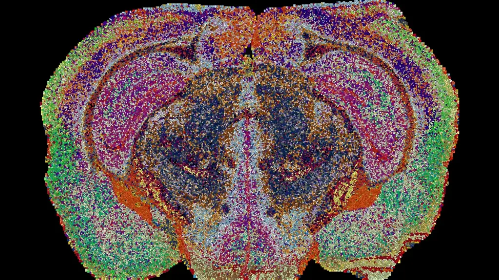
To bridge this divide, the EPFL team employed mass spectrometry imaging—a sophisticated technique that allows for the localized identification of molecules across thin slices of biological tissue. By using a laser to scan each slice with micron-level precision, the researchers could pinpoint exactly which lipids were present at any given coordinate. By processing 109 brain slices from 11 mice, the team generated a staggering 7 million measurements. These data points were then synthesized into a robust 3D reconstruction, powered by bespoke machine learning algorithms designed specifically to handle the complexity of lipidomic data.
Identifying 539 Distinct Chemical Zones: The "Lipizones"
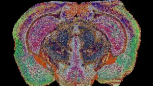
The resulting Lipid Brain Atlas provides a panoramic view of the brain that is as organized as it is unexpected. The mapping revealed that lipids are not randomly dispersed; rather, they are arranged in highly specific, recognizable patterns. The researchers identified 539 chemically distinct territories, which they have dubbed "lipizones." Each of these zones possesses a unique combination of lipids, creating a distinct chemical signature that allows researchers to identify the origin of a brain tissue sample simply by analyzing its lipid profile.
Many of these lipizones align perfectly with established anatomical structures or known cell types, reinforcing the validity of the mapping. However, the atlas also uncovered surprising relationships between disparate parts of the brain. Some regions, while physically separated by anatomy, shared identical chemical signatures, suggesting they may be linked by functional requirements that transcend their spatial location.
"In effect, lipids work like postal codes, giving distant but related parts of the brain the same chemical address," says D’Angelo. This discovery challenges the traditional reliance on gene activity as the sole indicator of regional identity. While genetic maps are invaluable, the lipid atlas suggests that the brain’s chemical organization offers an additional, vital layer of information regarding how different areas of the brain are functionally related and organized.
Hidden Chemical Patterns and the Complexity of White Matter
One of the most striking revelations of the project concerns the nature of white matter. Traditionally, white matter—the tissue containing the axons that connect various parts of the brain—has been viewed as a relatively uniform "highway" system. However, the Lipid Brain Atlas shows that this is a misconception. Instead of being chemically consistent, white matter is composed of numerous distinct lipid zones, each with its own unique composition.
"This shows that the cells which make myelin are far more varied in their chemistry than their genes alone would suggest," notes Luca Fusar Bassini. The diversity of lipids within white matter suggests that the support system for nerve fibers is much more specialized than previously assumed. Furthermore, the team identified previously unrecognized chemical boundaries within the choroid plexus, the specialized tissue responsible for the production of cerebrospinal fluid, and in the linings of the brain’s internal cavities. These findings suggest that the brain’s chemical architecture is far more granular and specialized than either anatomy or genetics could reveal on their own.
Pregnancy and the Brain’s Adaptive Chemistry
Beyond static mapping, the atlas provided an unprecedented look at how the brain’s chemical composition fluctuates in response to physiological states, specifically during pregnancy. By comparing the lipid maps of pregnant mice to those of non-pregnant subjects, the researchers observed widespread, significant shifts in lipid distribution throughout the brain.
These changes were particularly pronounced in the cortex, the brain’s outer layer responsible for higher-order functions. Additionally, the team observed a sharp increase in galactosyl ceramide—a crucial component of myelin—within the white matter. These shifts were found to be more extensive than the differences typically observed between male and female brain lipid profiles.
These findings suggest that the brain is not a static organ, but a dynamic one capable of "retuning" its own chemical wiring to meet the physiological demands of pregnancy. "The Atlas reveals that the brain constantly adjusts its lipids to meet the body’s changing needs," explains Gioele La Manno. This adaptive capacity highlights the importance of lipid metabolism in neural plasticity and suggests that the brain’s chemical state is a reflection of the body’s overall health and physiological status.
A New Era for Neurobiological Research
The implications of this atlas extend far beyond the laboratory in Lausanne. The Lipid Brain Atlas has been made freely available online, inviting scientists worldwide to explore the previously inaccessible chemical landscape of the brain. By integrating this lipidomic data with existing genetic and cellular atlases, researchers now have a multi-dimensional tool to investigate the mechanisms of brain aging, development, and pathology.
For researchers studying conditions such as Alzheimer’s, depression, or developmental neurobiology, the atlas provides a much-needed baseline of "healthy" brain chemistry. By identifying the chemical markers that define a healthy brain, scientists can now more easily pinpoint the specific lipid disruptions that may precede or accompany disease states.
While the current map is derived from mouse models, the methodology developed by the EPFL team is highly translatable. The primary contribution is not merely the mouse map itself, but the demonstration that the brain possesses a highly organized, predictable chemical map—and the establishment of a robust framework to uncover these patterns in other species, including humans.
"What transfers to humans isn’t the map, it’s the method, and the fact that we now know that a healthy brain has a very organized lipid map at all," says Fusar Bassini. As the field of lipidomics continues to mature, this atlas serves as a foundational resource, signaling a new era in which the brain’s "fatty" composition is no longer a hidden variable, but a central component of our understanding of the most complex organ in the body.