





Why do some new skills seem to click almost instantly, feeling intuitive from the very first attempt, while others remain frustratingly out of reach despite hours of diligent practice? For decades, the conventional wisdom surrounding mastery has focused almost exclusively on the brain’s cognitive capacity, the intensity of individual effort, or the nebulous concept of innate talent. However, emerging research from Tohoku University suggests that the secret to lasting learning may lie in an often-overlooked factor: the internal physiological state of the body and its constant, rhythmic dialogue with the brain.
In a study published on August 25, 2026, in the journal iScience, researchers have demonstrated that the vagus nerve—a sprawling communication highway that links our internal organs to the central nervous system—plays a critical role in cementing motor skills. By stimulating this nerve in mice immediately following practice sessions, the research team successfully strengthened long-term learning, proving that the body’s physiological "environment" can be tuned to make the brain more receptive to durable, lasting change.
The Vagus Nerve: A Gateway to Plasticity
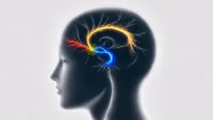
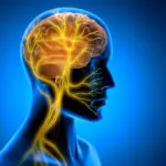
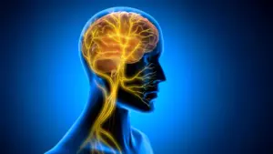
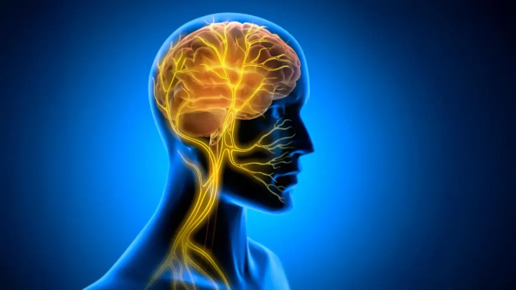
The vagus nerve serves as one of the most vital conduits in the human body. As a primary component of the autonomic nervous system, it acts as a two-way information highway, carrying sensory data from the heart, lungs, and digestive tract up to the brain, while simultaneously transmitting regulatory instructions from the brain back down to these internal organs. Because of its expansive reach and its influence over various bodily functions, the vagus nerve has long been a target for medical intervention.
Vagus nerve stimulation (VNS) is already a clinically approved therapeutic technique used to treat several neurological and psychiatric disorders, including refractory epilepsy and depression. Historically, scientists have viewed VNS primarily as a form of neuromodulation—a way to alter the activity of neurotransmitter systems such as norepinephrine and acetylcholine. By triggering these systems, VNS was thought to "prime" the brain for better function.
However, the team at Tohoku University, which specializes in super-network brain physiology, suspected that the mechanism behind VNS-enhanced learning might be more nuanced than previously realized. They hypothesized that the benefits of stimulating this nerve might involve physical, rhythmic changes in the blood vessels within the brain, thereby altering the metabolic environment in which memory consolidation occurs.
The Experimental Framework
To test this hypothesis, the researchers developed a specialized, miniaturized cuff electrode capable of being permanently attached to the left cervical vagus nerve in mice. With this technology, they were able to deliver precise stimulation while the mice engaged in a specific motor task: the horizontal optokinetic response (HOKR).
The HOKR is a classic cerebellum-dependent eye movement task. In this experiment, mice were taught to improve their ability to track moving visual stripes. This task is remarkably similar to the natural, automatic stabilization movements humans perform when standing on a stationary platform and watching a high-speed train pass by; the brain must learn to coordinate visual input with precise motor adjustments to keep the eyes locked on a target.
Crucially, the researchers altered the timing of the intervention to see if the physiological "timing" of stimulation mattered as much as the stimulation itself. Instead of delivering VNS during the actual learning task—when the brain is busy processing sensory input and attempting to execute movements—the team applied the stimulation only after each training session had concluded.
A Hidden Window for Memory Consolidation
The results were striking. The stimulation provided no immediate performance boost while the mice were in the midst of training. However, the benefits became significantly apparent in the days that followed. Mice that received post-training VNS demonstrated superior long-term retention of the task compared to the control groups.
This temporal pattern suggests that VNS does not necessarily aid in the initial acquisition of a skill but rather bolsters the post-training "offline" period. This is the critical window when the brain is hard at work consolidating new information, transforming fragile, short-term practice into durable, long-term memory.
"The key point is that VNS was delivered only after training," explains Professor Ko Matsui, a lead researcher on the project. "Our findings suggest that VNS may open a hidden window of opportunity for enhanced learning by making the brain environment more receptive to long-lasting change."
Rhythmic Vascular Dynamics: The Missing Link
To uncover the physiological mechanism driving these results, the team employed fiber photometry to monitor blood volume activity within the cerebellar flocculus—a region of the brain that is fundamentally involved in HOKR learning.
The researchers discovered that a single round of VNS triggered a distinct two-phase vascular response: an initial, brief decrease in local blood volume followed by a subsequent, more sustained increase. When VNS was applied repeatedly, these changes in blood volume evolved into rhythmic oscillations.
The data indicated a clear correlation between these vascular rhythms and the animals’ learning outcomes. Mice that exhibited larger and more pronounced blood volume oscillations during the post-training window performed significantly better on the task by the fifth day. This discovery suggests that the brain’s vascular environment—its blood flow and metabolic supply—is not a passive bystander but an active participant in the creation of long-term memory. By modulating these rhythms, the body effectively signals to the brain that the recently practiced information is of high value and worthy of being "saved" permanently.
"Our brains may be more strongly influenced by the body than we imagine," says lead author Junyu Chen. "By tuning the brain’s metabolic environment, including rhythmic vascular movements, we may eventually unlock capacities that would otherwise remain latent."
Implications for the Future of Learning
This research marks a significant shift in how scientists view the brain-body connection. If the metabolic environment of the brain can be actively manipulated to improve the retention of motor skills, the implications for human health and education are profound.
While the current study was confined to mouse models, the findings provide a robust framework for future human research. The ability to enhance plasticity through non-invasive or minimally invasive stimulation could eventually be used to accelerate recovery for stroke patients, assist those with neurodegenerative diseases in retaining motor function, or even provide new strategies for rapid skill acquisition in high-stakes fields.
The research team at Tohoku University is now turning its attention toward refining these stimulation protocols. The next phase of their work will focus on determining the optimal frequency and duration of stimulation, as well as mapping the precise pathways through which the body communicates these signals to the brain to support long-term plasticity.
As scientists continue to pull back the curtain on the complex, two-way dialogue between our organs and our neural networks, it is becoming increasingly clear that learning is not a purely cerebral endeavor. Instead, it is a holistic process, deeply rooted in the rhythmic, physiological symphony of the entire body. By understanding how to harmonize this internal conversation, researchers are moving closer to unlocking the full, often latent potential of the human brain.