






Why do some skills seem to click almost instantly, embedding themselves into our muscle memory with minimal effort, while others remain frustratingly elusive despite hours of diligent practice? For years, educators and neuroscientists have attributed this disparity to a combination of innate talent, individual motivation, and the volume of repetition. However, new research suggests that the secret to mastery may lie in a biological mechanism that has little to do with raw intellect or sheer willpower. Instead, the difference may come down to whether the brain is in the optimal physiological condition to transform fleeting practice into a permanent, lasting memory.
The latest findings, published in iScience on August 25, 2026, suggest that learning is not a process that occurs in isolation within the cranium. Rather, it is a whole-body endeavor. Researchers at Tohoku University, specializing in the physiology of the brain’s complex neural networks, have demonstrated in mice that the body’s internal organs play a critical, previously overlooked role in solidifying motor skills. By stimulating the vagus nerve—a primary communication highway between the body and the brain—immediately following a training session, the team was able to significantly strengthen long-term motor learning. This discovery challenges the long-held view of the brain as a solitary command center, suggesting instead that the body actively regulates the brain’s ability to "save" new information.
The Vagus Nerve: A Gateway to Neural Plasticity
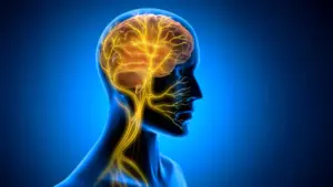
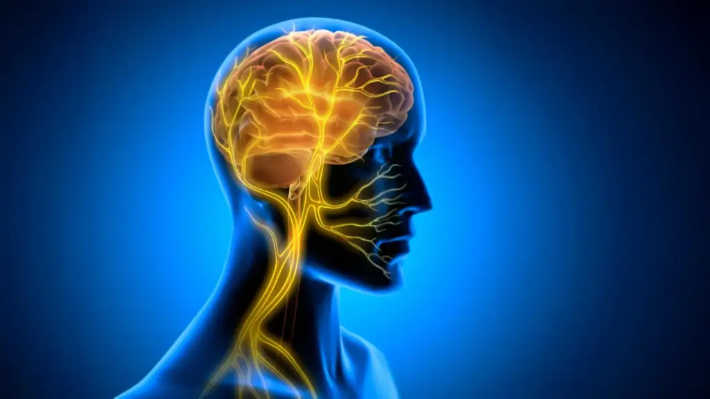
The vagus nerve is one of the most vital components of the autonomic nervous system. Acting as a bidirectional information superhighway, it constantly ferries sensory data from the heart, lungs, and digestive tract up to the brainstem, while simultaneously carrying regulatory signals from the brain back down to these organs. Because of its expansive reach, the vagus nerve has long been a target for medical intervention. Vagus Nerve Stimulation (VNS) is already an established clinical therapy, approved for the treatment of various neurological and psychiatric conditions, including refractory epilepsy and depression.
Historically, scientists have viewed VNS through the lens of neuromodulation, focusing on how electrical pulses can alter the activity of neurotransmitter systems—the chemical messengers that facilitate communication between neurons. However, the Tohoku University study proposes a fundamentally different, perhaps more mechanical, mechanism. The researchers hypothesize that VNS influences learning by inducing rhythmic changes in the blood vessels within the brain. By modulating the vascular environment, the vagus nerve may be creating a metabolic "window of opportunity" that allows the brain to consolidate memories more effectively.
To put this hypothesis to the test, the team developed a specialized, miniaturized cuff electrode, which they attached to the left cervical vagus nerve in mice. This allowed for precise control over the stimulation process. The researchers then monitored the mice as they engaged in a horizontal optokinetic response (HOKR) task. HOKR is a cerebellum-dependent learning exercise that requires subjects to improve their ability to track moving visual stripes. The task serves as an excellent model for motor learning; it mimics the automatic, reflexive eye movements a human makes when standing on a station platform and watching a train pass by.
The Critical Timing of Post-Training Stimulation
One of the most revealing aspects of the study was the timing of the intervention. The researchers applied VNS only after each training session had concluded, rather than during the actual performance of the HOKR task. Interestingly, the mice showed no immediate improvement in their performance while they were undergoing training. The stimulation did not act as a "performance enhancer" in the moment, nor did it make the act of tracking the stripes easier while the training was in progress.
Instead, the true benefits of the intervention manifested in the days that followed. Mice that received VNS after their training sessions demonstrated significantly stronger, more durable long-term learning compared to the control group. This delayed effect provides a vital clue into the nature of memory formation. It suggests that the vagus nerve does not necessarily help the brain acquire information, but rather helps the brain "consolidate" it. Consolidation is the post-training process during which newly acquired information is stabilized and integrated into existing neural networks, transforming a fragile, temporary trace into a durable, long-term memory.
Professor Ko Matsui, a lead researcher on the project, emphasized the importance of this temporal distinction. "The key point is that VNS was delivered only after training," Matsui explained. "Our findings suggest that VNS may open a hidden window of opportunity for enhanced learning by making the brain environment more receptive to long-lasting change." By intervening after the effort is expended, the stimulation appears to prime the brain to lock in the progress made during the practice session.
Rhythmic Vascular Movements and the Brain’s Metabolic Environment
To understand why this post-training stimulation led to such robust results, the team turned their attention to the physiological changes occurring within the brain itself. Specifically, they utilized a technique called fiber photometry to measure blood volume activity near the cerebellar flocculus—the specific area of the cerebellum responsible for coordinating the HOKR learning process.
The results revealed a sophisticated, two-phase vascular response following a single round of VNS. Initially, the local blood volume in the cerebellar flocculus decreased briefly, followed by a delayed rise. When the researchers repeated the stimulation, this sequence evolved into a series of rhythmic oscillations in blood volume. The team discovered a striking correlation: the mice that exhibited larger, more pronounced blood volume oscillations during the post-training period consistently demonstrated superior learning performance by the fifth day of the experiment.
This finding suggests that the vascular environment is not merely a passive provider of oxygen and nutrients to the brain, but an active participant in the learning process. The rhythmic pulsing of blood vessels may be facilitating the movement of metabolic waste or the delivery of essential signaling molecules, creating a more favorable environment for the synaptic changes that underlie memory. It is a striking example of "vascular tuning," where the body’s physical rhythms dictate the efficiency of the brain’s cognitive restructuring.
Lead author Junyu Chen noted the profound implications of these findings for our understanding of the brain-body connection. "Our brains may be more strongly influenced by the body than we imagine," Chen said. "By tuning the brain’s metabolic environment, including rhythmic vascular movements, we may eventually unlock capacities that would otherwise remain latent." This suggests that the limits of our learning ability may be partially defined by how well we can synchronize the physiological state of our body with the requirements of our brain.
Unlocking the Potential of the Brain-Body Axis
The implications of this research extend far beyond the laboratory. By identifying the specific biological pathways through which the body influences neural plasticity, the researchers have opened the door to new strategies for enhancing learning and rehabilitation. If VNS or other techniques can be used to "open" this window of opportunity for memory consolidation, it could revolutionize how we approach the treatment of motor disorders or even how we accelerate the acquisition of new skills in healthy individuals.
However, the team remains cautious and focused. The current study is an essential first step in mapping this complex, bidirectional communication pathway. Future research will be directed toward refining these stimulation protocols, determining the optimal frequency and duration of VNS to maximize its effects, and understanding the molecular signals that link vascular rhythms to long-term plasticity at the cellular level.
As the scientific community continues to peel back the layers of how the vagus nerve influences cognitive function, the hope is to gain a more comprehensive understanding of how learning becomes permanent. By studying the dialogue between the internal organs and the brain in greater detail, scientists are moving closer to answering the fundamental question of why some people—and some brains—are more adept at mastering new challenges. In the future, the difference between a frustrating plateau and a breakthrough in learning may be as simple as ensuring that the body is in the right rhythm to help the brain learn.