







Why do some new skills seem to click almost instantly while others remain frustratingly difficult, even after hours of focused, repetitive practice? For decades, scientists have attributed this disparity to individual talent, the intensity of effort, or the quality of instruction. However, emerging research from Tohoku University suggests that the secret to mastering a new skill may lie in a biological factor that has long been overlooked: the state of the brain’s internal environment during the post-training recovery phase.
A study published in the journal iScience on August 25, 2026, reveals that the body’s internal organs play a far more active role in consolidating memory and motor skills than previously understood. By leveraging the vagus nerve—a critical communication highway between the body and the brain—researchers have successfully demonstrated in mice that stimulating this pathway after practice can significantly strengthen long-term motor learning. This discovery shifts the focus of neuroscience away from purely cerebral processes and toward a more holistic view of the body-brain connection.
The Vagus Nerve as a Learning Catalyst
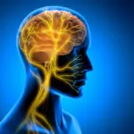
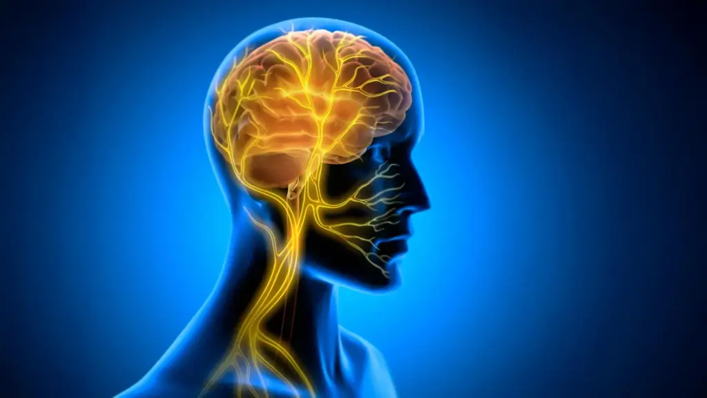
The vagus nerve is one of the most vital components of the autonomic nervous system. Acting as a major information highway, it facilitates a constant, bidirectional stream of signals between the brain and the body’s internal organs, such as the heart, lungs, and digestive tract. While the brain sends "top-down" instructions to these organs to regulate essential life functions, the nerve simultaneously carries "bottom-up" sensory information back to the brain.
Scientists have long been interested in modulating this pathway through Vagus Nerve Stimulation (VNS), a clinical technique that uses electrical impulses to influence neural activity. VNS is already an established therapeutic tool, approved for the treatment of several medical conditions, including epilepsy and treatment-resistant depression. Traditionally, the efficacy of VNS in these contexts has been explained through neuromodulation—the capacity for the stimulation to alter the activity of neurotransmitter systems, such as norepinephrine and acetylcholine, which are known to be involved in attention and mood regulation.
However, the team at Tohoku University, specializing in super-network brain physiology, hypothesized that VNS might be doing more than just adjusting neurochemistry. They suspected that the stimulation might be triggering mechanical or metabolic shifts within the brain’s vascular system, thereby creating a more favorable environment for learning to take root.
Rethinking the Timing of Stimulation
To test this theory, the research team developed a specialized, small-cuff electrode designed to be securely attached to the left cervical vagus nerve in mice. The researchers then monitored the mice as they engaged in a horizontal optokinetic response (HOKR) learning task. HOKR is a cerebellum-dependent eye movement task that requires mice to learn how to track moving visual stripes. This task is remarkably similar to the automatic eye movements a person experiences when standing on a platform and observing a train passing by, requiring precise coordination between the visual system and motor control.
A pivotal aspect of the study design was the timing of the VNS. Rather than applying the stimulation during the training sessions, the researchers delivered the impulses only after the training had concluded. This deliberate choice allowed the team to isolate the effects of stimulation on memory consolidation—the physiological process by which the brain stabilizes a memory trace after the initial acquisition of information.
The results were striking. The stimulation did not provide an immediate performance boost; the mice did not demonstrate superior eye movement coordination while they were actively practicing the task. Instead, the benefits emerged over time. On the days following the training, the mice that had received the post-training VNS exhibited significantly stronger long-term learning compared to the control group.
"The key point is that VNS was delivered only after training," explains Professor Ko Matsui, a senior researcher on the project. "Our findings suggest that VNS may open a hidden window of opportunity for enhanced learning by making the brain environment more receptive to long-lasting change." This suggests that the brain enters a unique physiological state after the learning process ends, and that the vagus nerve can act as a trigger to "lock in" those newly formed neural pathways.
The Role of Rhythmic Vascular Oscillations
Having observed the behavioral improvements, the researchers sought to uncover the underlying biological mechanism occurring within the brain. They focused their investigation on the cerebellar flocculus, a region of the brain critical for the HOKR learning task. Using fiber photometry, a technique that allows researchers to monitor neural and vascular activity in real-time, the team observed the brain’s internal response to the VNS pulses.
They discovered that a single round of VNS triggered a distinct, two-phase vascular response. Initially, the local blood volume in the cerebellar flocculus decreased, followed by a delayed rise. When the researchers repeated the VNS sessions, this response evolved into rhythmic oscillations in blood volume. These rhythms appeared to be directly correlated with the degree of learning retention. Mice that exhibited larger, more pronounced blood volume oscillations were found to be the most proficient at the task by the fifth day of the experiment.
This evidence suggests that the vagus nerve does not merely broadcast signals to neurons; it may also influence the brain’s metabolic environment by modulating blood flow. By creating a rhythmic vascular pulse, VNS may be optimizing the delivery of nutrients or the clearance of metabolic waste in areas of the brain undergoing plasticity, thereby facilitating the structural changes required for long-term memory storage.
"Our brains may be more strongly influenced by the body than we imagine," says lead author Junyu Chen. "By tuning the brain’s metabolic environment, including rhythmic vascular movements, we may eventually unlock capacities that would otherwise remain latent."
Future Implications for Brain-Body Science
The implications of this research are significant for the broader field of neuroscience. By demonstrating that the vagus nerve can be used to prime the brain for long-term memory consolidation, the study provides a new framework for understanding the interplay between bodily states and cognitive performance.
For scientists, the next phase of this research will involve refining the stimulation protocols to better understand the optimal timing, frequency, and intensity of VNS required to produce the most durable learning effects. Furthermore, the team aims to determine the precise molecular and cellular mechanisms that bridge the gap between rhythmic vascular changes and synaptic plasticity—the ability of connections between neurons to strengthen or weaken over time.
As researchers continue to probe this two-way communication pathway, the hope is that these insights will move beyond the laboratory and eventually inform new therapeutic strategies. While currently focused on motor learning in animal models, the study opens the door to a deeper understanding of how humans might one day enhance their own capacity for learning. By bridging the divide between the body’s physiological rhythms and the brain’s cognitive processes, this research represents a step forward in our comprehension of how we turn experience into lasting knowledge. The ability to "unlock" latent cognitive potential through non-invasive stimulation remains a distant but increasingly plausible goal, rooted in the realization that the brain does not learn in a vacuum, but rather as part of a complex, interconnected biological whole.