







Why do some new skills feel intuitive, clicking into place almost instantly, while others remain elusive despite hours of dedicated practice? For decades, scientists have debated the role of innate talent, individual effort, and cognitive capacity in the acquisition of expertise. However, a groundbreaking study from Tohoku University suggests that the secret to lasting learning may not be found within the brain’s neurons alone. Instead, it may rely on a critical dialogue between the brain and the body, mediated by one of the nervous system’s most vital communication pathways: the vagus nerve.
In research published in the journal iScience on August 25, 2026, scientists have demonstrated in mouse models that stimulating the vagus nerve immediately following a training session can significantly strengthen long-term motor learning. This discovery challenges the traditional view of the brain as a self-contained processor, highlighting a previously overlooked role for systemic body-to-brain signaling in the consolidation of new skills. By effectively "tuning" the brain’s internal environment, researchers believe they may have discovered a way to make the mind more receptive to permanent change.
The Vagus Nerve: A Superhighway for Learning
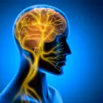
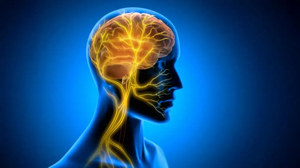
The vagus nerve is often described as the body’s information superhighway. It acts as a primary conduit, transmitting a constant stream of data from the internal organs to the brain while simultaneously carrying executive instructions from the brain back to those organs. Because of its expansive reach and its ability to modulate physiological states, the vagus nerve has long been a target for medical intervention. Vagus nerve stimulation (VNS)—a clinical procedure that uses electrical impulses to influence this pathway—is already an FDA-approved treatment for conditions such as epilepsy and treatment-resistant depression.

Historically, however, the scientific community has viewed VNS primarily as a tool for neuromodulation. The prevailing theory was that VNS worked by altering the activity of neurotransmitter systems, such as norepinephrine or acetylcholine, which are known to be involved in arousal and attention. The new study from Tohoku University adds a compelling new layer to this understanding: the possibility that VNS influences learning not just through chemical signals, but through rhythmic, mechanical changes in the blood vessels inside the brain itself.
To investigate this hypothesis, researchers developed a specialized, miniaturized cuff electrode capable of being permanently attached to the left cervical vagus nerve in mice. With this tool, the team could precisely control the timing and duration of electrical stimulation. They then set the mice to a task involving horizontal optokinetic response (HOKR) learning. This is a cerebellum-dependent motor task that requires the subject to improve its ability to track moving visual stripes. The response is a fundamental reflex, mirroring the way a human passenger on a platform might automatically track a passing train with their eyes to maintain focus.
The Critical Window After Practice
One of the most striking findings of the study was the importance of timing. The researchers deliberately applied VNS after each training session, rather than during the learning task itself. When the stimulation was delivered during the task, there was no immediate, observable improvement in performance. The mice did not suddenly become better at tracking stripes while the electricity was active.
However, the results on subsequent days told a very different story. The mice that received VNS in the post-training window demonstrated significantly stronger long-term retention of the skill. This temporal separation is crucial; it suggests that VNS does not necessarily aid the acquisition of information, but rather the consolidation of it. It appears that the stimulation acts upon the brain’s post-training state, potentially transforming a fleeting, fragile memory into a more durable, long-term neural structure.
Professor Ko Matsui, a key researcher on the project, emphasizes the significance of this observation. "The key point is that VNS was delivered only after training," Matsui notes. "Our findings suggest that VNS may open a hidden window of opportunity for enhanced learning by making the brain environment more receptive to long-lasting change." This implies that there is a biological state—a "window of receptivity"—that follows intense mental or physical effort, and that the vagus nerve acts as a gatekeeper to that state.
Rhythmic Changes in Brain Blood Volume
To understand how this stimulation translates into improved memory, the team delved into the physiological environment of the cerebellum, specifically the cerebellar flocculus—an area known to be critical for the coordination of eye movements. Using a sophisticated imaging technique called fiber photometry, the researchers monitored blood volume activity in the brain as it responded to the vagus nerve stimulation.
The data revealed that a single round of VNS triggered a distinct, two-phase vascular response. Initially, local blood volume within the cerebellar area decreased, only to be followed by a secondary rise after a short delay. When the stimulation was repeated over the course of the training program, this two-phase response evolved into rhythmic, recurring oscillations in blood volume.
Crucially, these vascular rhythms were not merely incidental; they were directly correlated with the mice’s ability to retain the skill. The researchers found that mice exhibiting larger, more consistent blood volume oscillations performed better on the HOKR task by the fifth day of the experiment. This provides strong evidence that the brain’s vascular environment is not a static background feature of cognition, but a dynamic participant in the learning process. By influencing these rhythms, VNS appears to be optimizing the metabolic environment, perhaps by facilitating the clearance of metabolic waste or by enhancing the delivery of nutrients during the period when the brain is "writing" new memories.
Implications for the Future of Neuroplasticity
The findings from Tohoku University offer a fresh perspective on the complex relationship between the body and the mind. For lead author Junyu Chen, the results serve as a reminder that cognitive function is deeply rooted in systemic physiology. "Our brains may be more strongly influenced by the body than we imagine," Chen says. "By tuning the brain’s metabolic environment, including rhythmic vascular movements, we may eventually unlock capacities that would otherwise remain latent."
The realization that the brain’s capacity for plasticity can be gated by peripheral nervous system signals opens up a host of possibilities for future research. Scientists are now looking to refine these stimulation protocols, moving toward a more precise understanding of how the frequency, timing, and intensity of VNS influence long-term neural changes.
If this "body-to-brain" dialogue can be fully mapped, it could lead to novel, non-invasive therapeutic approaches to enhance learning in both clinical and healthy populations. Whether it is helping patients recover motor functions after a stroke or finding ways to improve the speed at which individuals master complex new skills, the ability to "open" the brain’s window of opportunity is a tantalizing prospect.
For now, the study provides a vital piece of the puzzle. It shifts the focus from the brain as a lone master of learning to a more holistic view, where the rhythmic pulse of the vascular system, directed by the vagus nerve, plays a central role in transforming practice into permanent knowledge. As researchers continue to explore this two-way communication pathway, they hope to better understand not just how we learn, but how that learning becomes etched into our biology, providing a foundation for future advancements in human performance and cognitive health.