







In a significant advancement for neurobiology, researchers at the University of Birmingham have unveiled a promising new strategy to address harmful brain inflammation. By pinpointing a specific receptor that is already susceptible to existing pharmaceutical compounds, the study opens a viable path toward repurposing medications to treat a host of debilitating neurological and psychiatric conditions.
The research, published in the journal Brain and spearheaded by Professor Nicholas Barnes of the University of Birmingham’s College of Medicine and Health, focuses on the P2X7 receptor. Long suspected of playing a role in cellular signaling, this receptor has now been identified as a critical driver of neuroinflammation. By effectively blocking this mechanism, the research team demonstrated a significant reduction in inflammatory responses within human brain tissue, offering a potential lifeline for conditions ranging from traumatic brain injury (TBI) to Alzheimer’s and Parkinson’s disease.
Blocking a Key Driver of Brain Inflammation
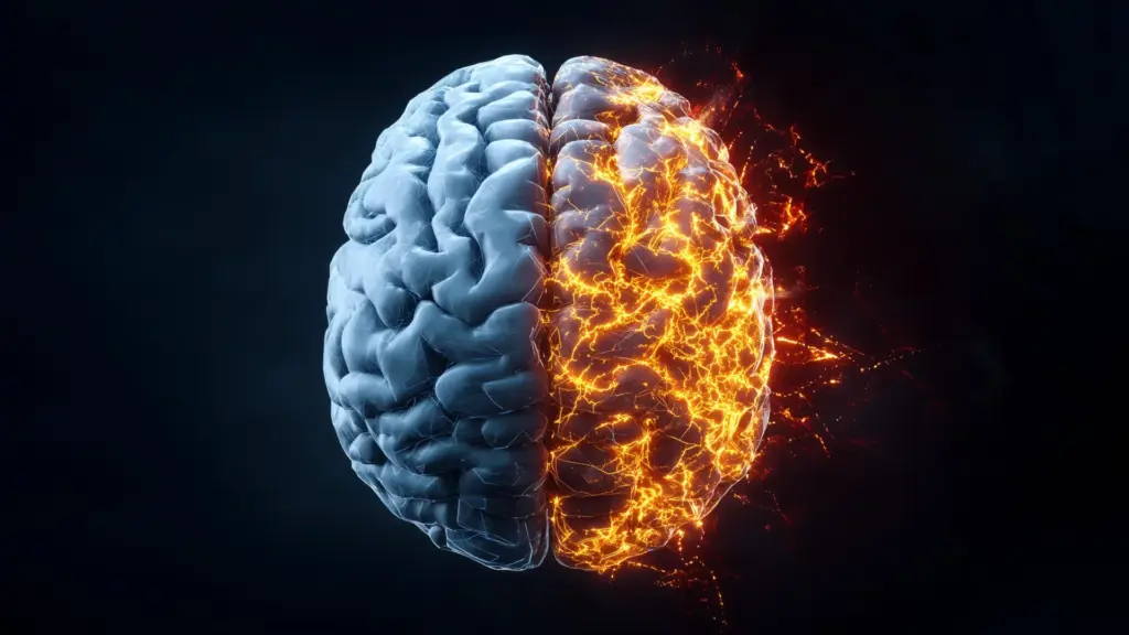
Neuroinflammation is a complex and often destructive process characterized by the activation of the brain’s immune system. While intended to be a protective response to injury or infection, chronic or unregulated inflammation can lead to the degradation of neural tissue, contributing to the progression of various diseases. For years, scientists have sought to understand the molecular "switches" that trigger this process.
The research team concentrated their efforts on the P2X7 receptor, which is known to facilitate the release of cytokines. Cytokines are a broad category of small proteins that act as chemical messengers, regulating the body’s inflammatory responses. When the P2X7 receptor is activated, it prompts an excessive release of these proteins, which can lead to a cascade of inflammation that damages healthy neurons.
To test the efficacy of inhibiting this receptor, the team employed a two-pronged approach. They utilized live cultures of human brain cells and, crucially, slices of human brain tissue obtained during necessary neurosurgical procedures. By introducing a specific antagonist—a substance that binds to a receptor to block its activity—the researchers observed a substantial drop in the inflammatory signaling pathways.
Professor Nicholas Barnes emphasized the potential impact of these findings. "This exciting discovery marks a major step toward repurposing existing therapeutics to combat neuroinflammation at its source," Barnes stated. "The identification of this receptor could have far-reaching implications for some of the most debilitating and widespread brain disorders such as Alzheimer’s Disease, Parkinson’s and Multiple Sclerosis, or inflammation-linked psychiatric conditions like schizophrenia and depression."
Studying the Brain’s Immune Cells
Central to the research is the role of microglia, the resident immune cells of the central nervous system. These cells act as the brain’s first line of defense, patrolling the neural environment and coordinating responses to trauma or cellular damage. However, when microglia remain in a chronically "activated" state, they contribute to the very inflammation they are meant to resolve.
Studying human microglia has historically presented a formidable challenge to neuroscientists. Once removed from the unique chemical and physical environment of the brain, these cells typically lose their defining characteristics and functional capabilities, making them difficult to study in a laboratory setting. To circumvent this, the Birmingham team developed a sophisticated, novel method for generating microglia-like cells from human peripheral monocytes—a type of white blood cell found in the bloodstream.
This innovative conversion process mimics a cellular transformation that researchers have recently identified as occurring naturally in the human brain during the aging process. By starting with blood samples, the researchers were able to create a robust and scalable platform for studying human microglial biology.
"Studying human microglia has long been a major challenge: once removed from their native brain environment, they rapidly lose their defining characteristics, likely due to the absence of critical regulatory signals," Professor Barnes explained. "Our approach involved the use of monocyte-derived microglia which provide a powerful, scalable, and virtually unlimited platform for studying human microglial biology with unprecedented precision."
By observing these cells, the team was able to witness the inflammatory signaling process in real-time. When the microglia became stressed, damaged, or neared the end of their lifecycle, they released signals that propagated inflammation. When the researchers introduced the P2X7 receptor antagonist, they successfully interfered with this signaling chain, effectively "quieting" the inflammatory response of the cells.
From Lab Models to Human Brain Tissue
While laboratory-grown models are essential for identifying molecular pathways, the true test of any potential therapy is whether it functions within the context of human biology. Having established a clear mechanism in their monocyte-derived microglia, the team proceeded to validate their findings using actual human brain tissue.
The tissue, provided through ethical protocols following neurosurgical procedures, allowed the researchers to observe how the P2X7 antagonist performed in a complex, multi-cellular environment. The results were consistent with the laboratory findings, showing that the receptor is a consistent and actionable target for mitigating inflammation across different human samples.
The success of this translation from laboratory models to clinical-grade human tissue is a vital milestone. It bridges the gap between fundamental research and clinical application, providing a strong evidentiary basis for further study. The research team is now looking toward the next phase: the development of clinical trials.
The need for such interventions is acute. For many patients suffering from neurodegenerative diseases or traumatic brain injuries, there are currently no effective pharmacological treatments that successfully target and reduce neuroinflammation to prevent long-term damage. The identification of a receptor that is already targeted by existing, well-understood drugs significantly shortens the typical timeline for drug development, as the safety profiles of these compounds have already been vetted in other clinical contexts.
"Having identified the response in the human monocyte-derived microglia, this provided the impetus to translate these findings with human brain obtained following neurosurgical procedures," Professor Barnes noted. "This successful translation means the next stage for this research is the development of clinical trials in patients with neurodegenerative conditions and patients with TBI where there are no effective pharmacological treatments to reduce the neuroinflammation and arising damage."
As the scientific community looks toward the future, this study offers a new framework for addressing the underlying drivers of brain health decline. By shifting the focus from simply managing symptoms to addressing the source of inflammatory signaling, the team at the University of Birmingham has illuminated a path that may eventually lead to therapies that protect the brain, preserve cognitive function, and improve the quality of life for millions of individuals living with chronic neurological conditions. The next steps will involve rigorous clinical testing to determine the most effective ways to leverage this discovery in human patients, potentially ushering in a new era of neuro-immunology treatment.