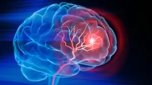








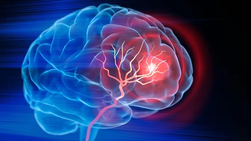
Biomedical engineers at Duke University have achieved a significant milestone in regenerative medicine, developing an injectable biomaterial that shows promise in helping the brain recover from the debilitating effects of an ischemic stroke. In a study published in the journal Cell Biomaterials, researchers demonstrated that by transforming the cavity left behind by lost brain tissue into a supportive, bioactive environment, they could recruit the body’s own immune system to facilitate repair, encourage vascularization, and restore motor function in mouse models.
The implications for this technology are vast. Ischemic strokes, which occur when a blood clot obstructs the flow of oxygen-rich blood to the brain, affect millions of people annually. While current emergency protocols—such as the administration of thrombolytic "clot-busting" drugs or surgical interventions to mechanically remove obstructions—are highly effective at saving viable tissue by restoring circulation, they are inherently limited. Once a region of the brain has succumbed to ischemia and the tissue has died, these standard treatments cannot reverse the damage.
The resulting void, or "stroke cavity," represents one of the most significant challenges in neurology. Currently, recovery for stroke survivors relies heavily on physical and occupational therapy. These rehabilitation methods are vital for teaching the surviving, healthy brain circuits to adapt and compensate for the lost functions, but they do not address the structural damage itself. The brain, unlike some other organs in the body, possesses a notoriously limited capacity for self-repair, often leaving patients with permanent neurological deficits.

A New Approach to Brain Engineering
"Once brain tissue has been lost, restoring blood flow is no longer enough," explains Tatiana Segura, the Robert Plonsey Distinguished Professor of Biomedical Engineering at Duke University and a lead figure in this research. "Our goal is to engineer the injured space so that immune, vascular, and neural repair processes can begin to work together."
To achieve this, Segura and her team moved beyond the concept of simply filling a hole. Instead, they aimed to create a scaffold that acts as a sophisticated, temporary "nursery" for the brain’s own cells. The team utilized MAPS—microporous annealed particle scaffolds. These are composed of individual, biocompatible hydrogel microparticles that, when injected into the cavity, self-assemble into a porous, sponge-like structure. This open architecture is critical; it provides a framework that allows cells to infiltrate, migrate, and establish the networks necessary for tissue remodeling.
The innovation lies not just in the material, but in how it is "programmed" to guide cellular behavior. The researchers focused their efforts on astrocytes, the versatile, star-shaped cells that serve as the support staff of the central nervous system. When the brain suffers an injury, astrocytes are among the first to respond. They communicate with the broader cellular environment by releasing extracellular vesicles (EVs)—minuscule lipid-bound packages filled with proteins, lipids, and genetic material. These EVs function as chemical messengers, instructing neighboring cells on how to react to the injury.
Keeping Repair Signals Where They Are Needed
Recognizing the potency of these natural signals, the Duke team harvested EVs from lab-grown astrocytes and integrated them into their scaffold system. However, they faced a hurdle: if injected directly into the brain, these signals would likely disperse or be degraded before they could have a meaningful therapeutic effect.
To overcome this, the researchers chemically tethered the EVs to the surfaces of the hydrogel microparticles. This strategy ensures that the signaling molecules remain concentrated within the porous scaffold, creating a high-density "instructional zone" for any immune or neural cells that migrate into the area. By doing so, the team effectively transformed the scaffold from a passive filler into an active, localized command center.
"We are not simply placing a material into the brain," Segura said. "We are engineering a local environment that can coordinate several parts of the repair response."
Through rigorous testing, the researchers identified a specific cocktail of signaling molecules—most notably IL-4 and C1q—that proved particularly adept at attracting beneficial immune cells to the site of the injury. When these signals were present, they successfully recruited macrophages and, surprisingly, a population of neutrophils that remained at the site for an extended period.
Challenging the Role of Neutrophils
The inclusion of neutrophils in the healing process was an unexpected finding that challenges long-held medical dogmas. Traditionally, neutrophils are viewed as the "first responders" of the immune system; they are often associated with the early, inflammatory stages of a stroke, where they can sometimes contribute to secondary tissue damage.
However, the data from the Duke study suggests that the function of neutrophils is highly dependent on context. When these cells were recruited by the MAPS scaffold in the later stages of the injury, they appeared to pivot from an inflammatory role to a regenerative one. When the researchers experimentally depleted this neutrophil population, the results were telling: the formation of new blood vessels, or angiogenesis, dropped significantly, and the scaffold itself underwent much less structural remodeling.
"This result changes how we think about neutrophils after stroke," said Shangjing Xin, the study’s lead scientist and a postdoctoral fellow in the Segura Laboratory. "Their role appears to depend on when they arrive, where they are located, and the signals they receive from their surroundings. Our study demonstrates a potential engineering strategy to recruit and retain these cells at the right time."
From Biological Repair to Functional Recovery
The biological success of the scaffold was confirmed by observing the physical state of the tissue within the cavity. As the immune cells populated the scaffold, the researchers observed the growth of new blood vessels, providing the necessary infrastructure for tissue survival. Furthermore, they detected an increase in axonal fibers—the long, thread-like projections of neurons that transmit electrical signals—extending into the previously hollow region.
The ultimate validation, however, was in the physical behavior of the mice. In a grid-walking test designed to assess motor coordination and precision in forelimb placement, the mice treated with the optimized scaffold showed marked improvement. By the eight-week mark, their performance had reached a level that was statistically indistinguishable from healthy control animals, a recovery that persisted for the duration of the study.
Importantly, the researchers conducted control experiments to see if the EVs could achieve these results on their own. They could not. When the EVs were administered without the MAP scaffold, the therapeutic effects—specifically the blood vessel repair—were significantly diminished. This proved that the porous architecture of the scaffold, coupled with the localized concentration of signaling molecules, was essential to the healing process.
The Path Toward Clinical Application
While these findings represent a significant leap forward, the research remains in the preclinical phase. The current studies have been limited to mouse models, and the material was injected directly into the brain, a procedure that requires further safety and feasibility assessments before it can be considered for human patients. Furthermore, the EVs currently used in the study are derived from rat astrocytes. The team is now moving toward using human induced pluripotent stem cell-derived astrocytes, which would offer a more scalable and clinically relevant source of signals, potentially allowing for greater control over the molecular "instructions" provided to the brain.
As the team looks to the future, they remain focused on the broader philosophy of their approach. The goal is not to "rebuild" the brain from scratch, but to restore the ecological balance of the injured tissue.
"You do not restore an ecosystem simply by containing the initial damage," Segura said. "You have to create the conditions that allow life to return. That is how we think about the stroke cavity. The material is not intended to reproduce the brain itself, but to create an environment where the body’s own cells can enter, communicate, and participate in rebuilding vascularized tissue."
By shifting the focus from simply preventing cell death to actively fostering a regenerative environment, the Duke team has opened a new frontier in stroke research—one that may one day allow doctors to do more than just manage the aftermath of a stroke, but to actively help the brain heal itself.