








A groundbreaking examination of an Alzheimer’s patient’s brain has provided some of the most compelling evidence to date that clearing amyloid plaques can halt the subsequent cascade of damage that defines the disease. By observing two distinct biological environments within the same organ—one where amyloid was cleared and one where it persisted—researchers have gained a unique, high-resolution view of how anti-amyloid treatments may fundamentally alter the trajectory of Alzheimer’s disease.
The findings, presented at the 2026 Alzheimer’s Association International Conference and published simultaneously in the journal JAMA, describe an "unusually detailed" analysis of brain tissue from a patient who had participated in a long-term clinical trial. The study revealed that brain regions successfully cleared of sticky amyloid plaques showed minimal or no buildup of tau—the toxic protein associated with neurodegeneration and cognitive decline. In stark contrast, adjacent areas of the brain where amyloid remained stubbornly present exhibited high levels of tau and clear markers of cellular destruction.
This discovery offers rare, direct human evidence that the removal of amyloid plaques does more than just clean up debris; it may actually protect the brain from the secondary, more destructive processes that lead to memory loss. The results suggest that if amyloid can be cleared thoroughly and early, the progression of Alzheimer’s could potentially be significantly slowed.

"Seeing both disease patterns side-by-side in the same brain gave us a rare opportunity to understand how amyloid removal affects other proteins that contribute to Alzheimer’s disease," said co-senior author David Wolk, MD, co-director of the Penn Memory Center and director of the Penn Alzheimer’s Disease Research Center at the Perelman School of Medicine at the University of Pennsylvania. "The findings provide some of the clearest human evidence to date that anti-amyloid therapies may limit the accumulation of tau and slow the brain changes that lead to memory loss and cognitive decline."
The Interplay of Proteins in Alzheimer’s Disease
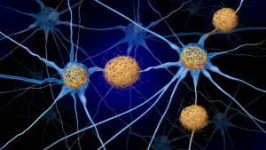
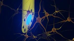
To understand the significance of this discovery, one must look at the two primary culprits in Alzheimer’s pathology: beta-amyloid and tau. In a healthy brain, these proteins serve various functions, but in the brains of those with Alzheimer’s, they become the agents of destruction. Beta-amyloid accumulates in the extracellular space—the area outside of nerve cells—where it forms dense, sticky deposits known as plaques. These plaques are widely considered the primary hallmark of the disease.
Tau, meanwhile, behaves differently. It typically accumulates inside the neurons themselves, forming tangled, fibrous structures. As these "tau tangles" proliferate, they disrupt the internal transport systems of the cells, eventually leading to the death of the neuron. This loss of neurons is the physical basis for the devastating symptoms of Alzheimer’s: memory loss, personality changes, and the eventual erosion of cognitive ability.
For decades, the medical community has operated under the hypothesis that amyloid buildup creates the environment necessary for tau to proliferate. However, confirming this link in a living human brain has been notoriously difficult. While clinical trials for anti-amyloid drugs have evolved rapidly—moving from the early, controversial approval of aducanumab to the more effective, fully approved treatments like lecanemab (Leqembi) and donanemab (Kisunla)—proving that removing the "trigger" (amyloid) prevents the "gunshot" (tau) has remained a major hurdle.
The primary barrier to this knowledge has been a lack of post-mortem brain data. Because few patients who undergo these novel anti-amyloid therapies donate their brains for exhaustive histological study, researchers have historically had to rely on PET scans or blood biomarkers, which, while useful, do not provide the granular, microscopic detail of direct tissue analysis.
A "Goldilocks" Scenario for Neuroscientific Discovery
The researchers at the University of Pennsylvania found their answer through a fortunate, albeit tragic, set of circumstances. The subject of the study was a man in his fifties who had been diagnosed with mild cognitive impairment caused by Alzheimer’s. He enrolled in a clinical trial for aducanumab and remained in the study for four and a half years, receiving 30 doses of the medication. He eventually passed away roughly four years after his final dose, more than a decade after the onset of his initial symptoms.
True to his commitment to science, the patient donated his brain, which was then analyzed by the Center for Neurodegenerative Disease Research in the Department of Pathology and Laboratory Medicine. What the team found was a startling, localized disparity in the brain’s condition.
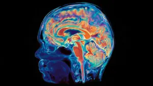
The brain’s folded surface consists of ridges, or "gyri," and the deep grooves between them, known as "sulci." The researchers discovered that the gyri had been effectively cleared of amyloid plaques and, consequently, contained very little tau. The sulci, however, had not responded as effectively to the treatment; they remained riddled with amyloid, and these same areas displayed significantly higher levels of tau tangles, chronic inflammation, and structural tissue degeneration.
"Many previous reports have shown either extensive amyloid clearance or limited clearance," noted co-senior author Edward B. Lee, MD, PhD, the John Q. Trojanowski, MD, PhD Professor for Neurodegenerative Disease Research. "This case represented a unique ‘Goldilocks’ scenario in which some regions cleared amyloid and others did not. It allowed us to directly compare what happened next in neighboring brain regions and better understand the relationship between amyloid, tau, and neurodegeneration."
Implications for Long-Term Brain Health
The discovery that amyloid clearance might offer a long-term "shield" against tau accumulation is a major development in the field of neurology. It suggests that the latest generation of monoclonal antibody therapies—which are designed to clear plaques—could have a ripple effect on the disease’s overall pathology that persists even after the drug has stopped being administered.
However, the researchers remain cautious about the limitations of the study. Because the investigation focused on a single individual, it cannot definitively establish the precise threshold of amyloid removal required to achieve lasting protection. Nor can it provide an absolute guarantee that the removal of amyloid is the sole driver of the reduced tau levels. Other biological factors, such as the brain’s internal environment or the patient’s individual genetics, could play a role in how different regions respond to therapeutic intervention.
Furthermore, the study highlights a discrepancy in how clinical success is measured. Most drug trials track patient progress over an 18-month window. This study suggests that the true, disease-modifying benefits of these medications—specifically the prevention of secondary tau-driven damage—might take years to fully manifest in a patient’s neurological profile.
Moving Toward Earlier Intervention
The overarching takeaway for the scientific community is the necessity of early intervention. If amyloid acts as a precursor that enables the development of more severe, tau-related damage, the window for effective treatment is likely much narrower than previously thought. Amyloid can accumulate in the brain for years, or even decades, before a patient ever experiences a moment of memory loss or cognitive confusion.
"Patients may accumulate amyloid for years before symptoms appear," said first author Christopher A. Brown, MD, PhD, an assistant professor of Neurology. "This case suggests that removing amyloid early may help limit the changes that ultimately damage brain cells. Ongoing trials will help determine whether starting treatment before symptoms begin can provide even greater benefit."
The research team is now focused on the next phase of investigation: identifying why certain brain regions are more receptive to amyloid-clearing treatments than others. By understanding the biological mechanics behind this regional variation, scientists hope to refine existing therapies to be more efficient and potentially more potent.
As the medical community continues to refine its approach to Alzheimer’s, this study stands as a critical piece of the puzzle. It moves the conversation beyond simply removing plaques and toward a broader goal of neuro-preservation. By confirming that the clearing of one protein can prevent the proliferation of another, the research provides a tangible, optimistic roadmap for future clinical trials—one where the focus shifts toward aggressive, early intervention to stop the cycle of destruction before it reaches the point of no return.
The study was supported by a series of grants from the National Institutes of Health, reflecting the high priority placed on uncovering the mechanistic relationship between the proteins that drive the world’s most common form of dementia. For patients and families navigating the uncertainty of an Alzheimer’s diagnosis, this evidence provides a meaningful glimpse into a future where the disease’s progression might be significantly altered.