







For patients living with severe, treatment-resistant depression, the path to recovery is often fraught with frustration. When traditional psychopharmacology and psychotherapy fail to provide relief, clinicians are increasingly turning to experimental interventions like deep brain stimulation (DBS). Now, a significant leap forward in this field has been made by researchers at the Mount Sinai Health System, who have identified three specific white matter pathways in the brain that are essential for achieving long-term improvement in patients undergoing subcallosal cingulate deep brain stimulation (SCC DBS).
This discovery, published on October 1 in the American Journal of Psychiatry, marks a pivotal moment in the development of neuromodulation therapies. By moving away from purely anatomical targeting and toward a strategy known as connectome-based targeting, researchers believe they can significantly refine the precision and effectiveness of this life-altering procedure.
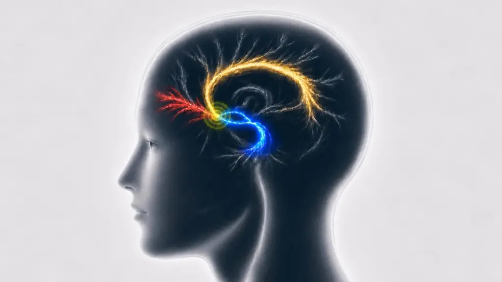
The research represents the largest multi-site study of its kind to date, aggregating data from several independent cohorts of patients who received SCC DBS. By analyzing these diverse groups, the researchers were able to confirm that lasting clinical improvements were consistently associated with electrical stimulation reaching three distinct pathways: the cingulum bundle, the forceps minor, and the medial uncinate fasciculus/ventral amygdalofugal pathways.
The Evolution of Surgical Targeting
Deep brain stimulation involves the surgical implantation of electrodes into specific regions of the brain. These electrodes deliver controlled electrical pulses designed to modulate the activity of circuits involved in mood regulation. However, as medical professionals have learned over decades of practice, the human brain is not a static map. The primary challenge in DBS has always been the precision of lead placement.
A common misconception is that stimulation only affects the immediate neurons surrounding the electrode. In reality, electricity travels along the brain’s white matter "highways," influencing larger, complex networks that may be located at some distance from the physical device. This explains a long-standing clinical enigma: why two patients with seemingly identical electrode placements can experience vastly different results. If one electrode successfully activates the necessary network and another does not—even if they are only millimeters apart—the clinical outcomes can range from total remission to no improvement at all.
Dr. Helen Mayberg, the Mount Sinai Professor in Neurotherapeutics and founding Director of the Nash Family Center for Advanced Circuit Therapeutics at the Icahn School of Medicine at Mount Sinai, emphasizes that individualized surgical planning is the new frontier of psychiatric treatment.
"This study underscores the importance of individualized surgical targeting for achieving optimal outcomes with deep brain stimulation for depression," Dr. Mayberg stated. "In any randomized controlled trial of a neuromodulation treatment, precise lead placement and confirmation that the intended brain target has been engaged are essential first steps. We still have much to learn about which patients are most likely to benefit from stimulation of this particular target, but developing imaging-based signatures that can match patients to their optimal treatment remains a central goal of our research."
Investigating the "Why" Behind Clinical Outcomes
To arrive at these findings, the team conducted a rigorous retrospective analysis. They utilized computer models to simulate the electrical fields generated by the implanted electrodes, effectively overlaying these fields onto high-resolution maps of individual brain connectivity. This allowed researchers to estimate exactly which white matter pathways were being stimulated in each patient and then cross-reference those patterns with the patients’ long-term clinical progress.
The data pool for this investigation was extensive, incorporating information from the BROADEN trial—a landmark clinical study—as well as data from the University of Toronto, where the foundational work for SCC DBS was originally pioneered.
The results were strikingly consistent. Across the various research centers, patients who experienced sustained improvements were those whose stimulation had successfully engaged the cingulum bundle, the forceps minor, and the medial uncinate fasciculus/ventral amygdalofugal pathways. Perhaps most telling was the observation that patients who failed to see results after two years of treatment shared a common characteristic: a consistent lack of engagement with the left cingulum bundle.
This finding suggests that the issue in non-responders may not be the treatment modality itself, but rather the specific "wiring" that was accessed. While the study did not go so far as to surgically move electrodes to prove this theory, it provides a compelling, evidence-based explanation for why some patients do not respond to standard positioning. It suggests that success in DBS is less about the "neighborhood" of the brain and more about the specific "circuits" being modulated.
Dr. Brian Kopell, Director of the Center for Neuromodulation at Mount Sinai, noted the profound implications of these findings for surgical practice. "This study addresses an important question in DBS: not simply which area of the brain to stimulate, but which connections within that area need to be engaged," Dr. Kopell said. "For a network disorder such as depression, millimeters can determine whether stimulation reaches the pathways we intend to modulate. Identifying and reproducibly engaging those pathways gives us a more precise surgical target and a framework for understanding why otherwise similar electrode placements may produce different clinical outcomes."
Toward a Future of Precision Psychiatry
The findings have already begun to influence current clinical research, most notably the Abbott TRANSCEND clinical trial. This randomized, controlled study is actively utilizing individualized brain connectivity maps to guide SCC DBS treatment, moving away from relying solely on anatomical landmarks in favor of patient-specific neuro-circuitry.
However, the researchers are careful to temper expectations, noting that while pathway engagement is a vital piece of the puzzle, it is not the only variable. The field must now determine how to identify which specific individuals are the best candidates for this invasive procedure and how to dynamically adjust stimulation parameters—such as amplitude, frequency, and pulse width—as a patient’s condition evolves over time.
One limitation noted in the study was the availability of imaging data. While some patient cohorts had individual diffusion MRI scans—which provide a detailed view of an individual’s unique white matter tracts—others did not. In those cases, the researchers had to rely on generalized connectivity maps. Encouragingly, the Emory University cohort allowed for a direct comparison between results derived from generalized maps versus those from individual scans, and both approaches yielded consistent findings. This reinforces the validity of the identified pathways, though it also points to the need for more widespread access to patient-specific imaging in future clinical settings.
Looking ahead, the team aims to delve deeper into the structural and functional integrity of these pathways. By characterizing abnormal brain states along these circuits, they hope to build predictive models that can guide clinicians in setting personalized stimulation parameters from day one.
"Our findings show precisely engaging these white matter pathways is a critical first step toward sustained response," said Dr. Ki Sueng Choi, an Associate Professor and Imaging Core Leader at the Center for Advanced Circuit Therapeutics at Mount Sinai, and the study’s corresponding author. "The next step is to characterize abnormal functional and structural brain states along these pathways to build more detailed models for setting patient-specific stimulation parameters, including amplitude, frequency, and pulse width, for precision medicine."
As this research continues, the goal remains clear: to transition from a "one-size-fits-all" approach to a model of precision medicine where neuro-imaging and surgical planning are inextricably linked. While further clinical research is required to fully quantify the impact of these methods, the Mount Sinai study offers a new, detailed map for navigating the complex circuits of the brain, bringing the prospect of reliable, lasting relief closer for those struggling with the most severe forms of depression.
The research was supported by the National Institutes of Health BRAIN Initiative, the Hope for Depression Research Foundation, and the DANA Foundation. Several of the study authors, including Dr. Mayberg, Dr. Kopell, and Dr. Choi, have disclosed consulting relationships with Abbott Laboratories, and some are named as co-inventors on patent applications related to the technologies used in the research. These disclosures underscore the deep involvement of both academic researchers and private industry in the pursuit of optimizing these advanced neuromodulation therapies.