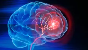






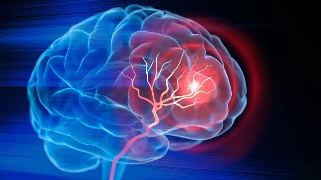
Biomedical engineers at Duke University have achieved a significant breakthrough in regenerative medicine, developing an injectable biomaterial designed to facilitate brain recovery following an ischemic stroke. In a study published in the journal Cell Biomaterials, researchers demonstrated that this novel material can transform the destructive cavity left behind by lost brain tissue into a highly favorable environment for healing. By recruiting the body’s own immune cells, fostering the growth of new blood vessels, and supporting the regeneration of neural tissue, the treatment led to measurable improvements in motor function in mouse models.
The Challenge of Repairing Brain Tissue After Stroke
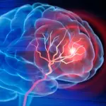
Ischemic strokes, which occur when a blood clot obstructs blood flow to the brain, remain a leading cause of long-term disability and death globally. Each year, millions of individuals are affected, facing the immediate threat of tissue death caused by oxygen deprivation. While modern emergency medicine—including clot-dissolving medications and mechanical thrombectomy—has revolutionized the ability to restore circulation and save viable tissue, these interventions are inherently limited.
Once brain cells die due to prolonged ischemia, the damage is essentially permanent. Standard clinical protocols focus on restoring perfusion to the remaining healthy tissue, but they do nothing to address the resulting physical void. Severe strokes often leave behind a cavity where healthy neural architecture once thrived. Currently, recovery for these patients depends almost exclusively on rehabilitation, which relies on the brain’s remaining circuits to adapt and compensate for lost function. While helpful, this process is indirect; it allows the brain to "work around" the damage rather than repairing it.
"Once brain tissue has been lost, restoring blood flow is no longer enough," said Tatiana Segura, the Robert Plonsey Distinguished Professor of Biomedical Engineering at Duke University. "Our goal is to engineer the injured space so that immune, vascular, and neural repair processes can begin to work together."
Building a Scaffold for Brain Repair
To address the void left by necrotic tissue, Segura and her research team sought to design a sophisticated environment that could stimulate multiple facets of the healing process simultaneously. The core of their strategy involves the use of microporous annealed particle scaffolds, known as MAPS.
These scaffolds are composed of individual hydrogel microparticles that, when injected, self-assemble into a highly organized, porous structure. This architectural design is critical; the open spaces created by the particles provide a three-dimensional framework that allows migrating cells to enter the injured site and utilize the scaffold as a foundation for rebuilding neural tissue.
Building upon the team’s prior success with this biomaterial, the current research focused on whether the scaffold could serve a dual purpose: acting as a structural bridge while simultaneously harnessing the body’s immune system to guide and accelerate the repair. To achieve this, the team targeted astrocytes. These star-shaped glial cells are fundamental to normal brain function, acting as both sentinels and support staff that respond rapidly to injury.
Astrocytes communicate with their environment and neighboring cells through the release of extracellular vesicles (EVs). These minute biological "packages" contain a complex cargo of proteins, lipids, and genetic material, acting as signaling molecules that can dictate the behavior of other cells. By capturing these vesicles, the researchers hoped to unlock a chemical key to brain regeneration.
Keeping Repair Signals Where They Are Needed
The challenge, however, lay in ensuring these signals remained at the site of the injury long enough to be effective. Injecting EVs directly into the brain would likely result in their rapid dispersion, rendering them ineffective. To circumvent this, the scientists chemically attached the EVs to the surfaces of the hydrogel microparticles within the MAPS scaffold.
This approach ensured that the healing signals remained highly concentrated within the damaged area, significantly increasing the probability that incoming immune cells would encounter these cues. "We are not simply placing a material into the brain," Segura explained. "We are engineering a local environment that can coordinate several parts of the repair response."
Through their experiments, the team identified a particularly potent combination of signaling molecules: IL-4 and C1q. These molecules proved remarkably effective at attracting specific immune cells to the scaffold, including macrophages and a persistent population of neutrophils.
An Unexpected Role for Neutrophils
The inclusion of neutrophils in the healing process was an unexpected, yet transformative, finding for the research team. Historically, neutrophils have been viewed through a negative lens in the context of stroke; they are often associated with acute inflammation and collateral tissue damage during the initial hours following an ischemic event.
However, the Duke study suggests that the role of these cells is far more nuanced. The research indicates that when neutrophils are present during the later stages of injury—and when they are directed by the specific signals and the micro-environment provided by the MAPS scaffold—they pivot from causing damage to facilitating repair.
To validate this, the researchers performed a depletion study, reducing the population of these immune cells at the injury site. The results were telling: when the neutrophils were removed, the formation of new blood vessels declined significantly, and the structural remodeling of the scaffold was hindered. This confirmed that these cells were essential, rather than detrimental, to the recovery process.
"This result changes how we think about neutrophils after stroke," said Shangjing Xin, the study’s lead scientist and a postdoctoral fellow in the Segura Laboratory. "Their role appears to depend on when they arrive, where they are located, and the signals they receive from their surroundings. Our study demonstrates a potential engineering strategy to recruit and retain these cells at the right time."
New Blood Vessels and Neural Growth
The biological impact of the scaffold extended beyond immune cell recruitment. As the immune cells populated the treated area, the researchers observed the formation of new, healthy blood vessels throughout the stroke cavity. Furthermore, there was a visible increase in axonal fibers—the essential neural pathways required for signal transmission—both within the cavity and in the surrounding brain tissue.
These physiological improvements translated into tangible functional gains for the mice. In grid-walking tests designed to evaluate motor skills and limb coordination, mice treated with the optimized scaffold showed significant recovery. By the eight-week mark, the treated mice exhibited performance levels that were statistically indistinguishable from those of healthy, non-injured control mice, a level of improvement that persisted throughout the remainder of the study.
Importantly, the researchers found that the scaffold itself was an essential component of this success. When they administered the EVs without the MAPS architecture, the restorative effects were absent. This finding suggests that the biomaterial does more than just transport therapeutic signals; its porous structure and ability to create a sustained, localized signaling environment are critical to the success of the repair response.
A Path Toward Clinical Translation
While the findings are promising, the researchers emphasize that this remains a preclinical approach. To date, the treatment has been tested exclusively in mouse models via direct injection into the brain. Significant hurdles remain before the technology can be considered for human application. Future research will need to rigorously assess the safety profile of the treatment, gain a deeper understanding of the interplay between different immune cell populations, and test the intervention in larger animal models that more closely replicate the complexity of human stroke.
Currently, the EVs used in the study are derived from primary rat astrocytes. As a next step, the Segura laboratory is working to utilize EVs produced by human induced pluripotent stem cell-derived astrocytes. This transition would not only provide a more scalable and clinically relevant source of material but also allow for greater precision in controlling the signaling cargo contained within the EVs.
As the team looks toward the future, the vision for this technology remains centered on the concept of restoration rather than simple containment. "You do not restore an ecosystem simply by containing the initial damage," Segura concluded. "You have to create the conditions that allow life to return. That is how we think about the stroke cavity. The material is not intended to reproduce the brain itself, but to create an environment where the body’s own cells can enter, communicate, and participate in rebuilding vascularized tissue."