







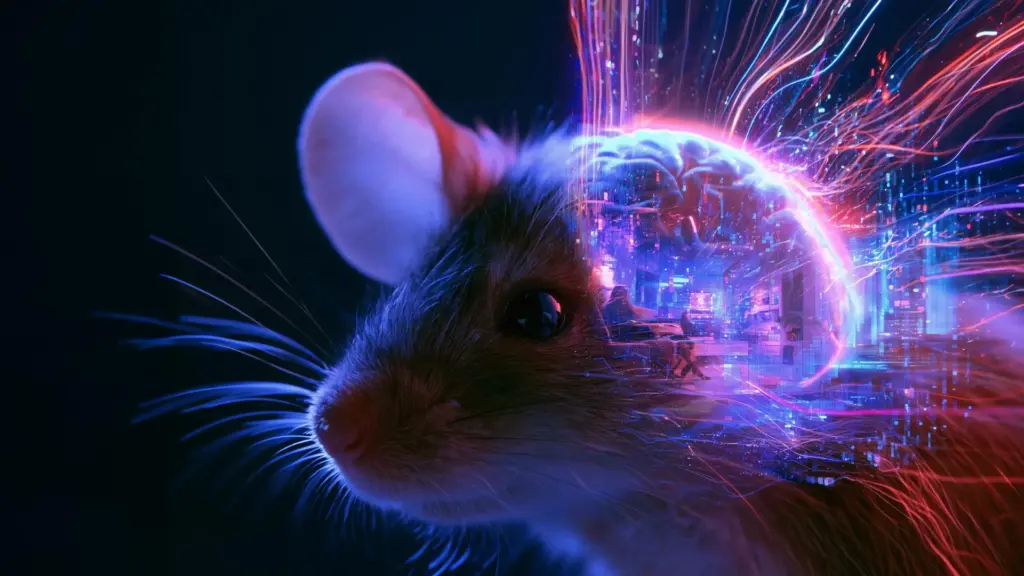
In a significant leap forward for neuroscience, researchers at University College London (UCL) have successfully reconstructed short, coherent videos using nothing but the raw neural activity recorded from the brains of mice. By effectively “watching” what the animals were seeing through the lens of their own visual cortex, the team has opened a new window into the complex mechanisms by which the brain translates light into an internal, subjective representation of the world.
The study, recently published in the journal eLife, represents a departure from traditional neuroimaging techniques. While previous research has attempted to decode human vision using functional magnetic resonance imaging (fMRI)—which captures broad blood-flow patterns—the UCL team opted for a higher-resolution approach. By focusing on the activity of individual neurons, the researchers have managed to capture a more granular and detailed picture of how visual information is encoded, filtered, and transformed within the biological circuitry of the brain.
Decoding the Language of the Visual Cortex
For decades, neuroscientists have grappled with the fundamental mystery of how the brain interprets the chaotic barrage of signals arriving from the retina. In human studies, researchers have long employed fMRI to monitor brain activity while subjects view movies or static images, attempting to map these patterns back to specific pixels on a screen. However, these methods often lack the precision required to understand the nuances of neural computation.
The UCL team, led by Dr. Joel Bauer of the Sainsbury Wellcome Centre at UCL, sought to bypass these limitations by examining the single-cell activity of mice. By monitoring individual neurons in the visual cortex, the researchers were able to observe the brain’s “language” in much higher fidelity.
“We wanted to have a better way of investigating how the brain interprets what we see,” Dr. Bauer explained. “The current methods of understanding what specific groups of neurons are representing are not very generalizable to situations which haven’t been specifically tested for. And so, we wanted to develop a method that can capture what is being represented in the brain and compare that to reality.”
This goal is central to the broader scientific pursuit of understanding the discrepancy between the physical world and the brain’s perception of it. The researchers are particularly intrigued by the differences between what is objectively present in front of an animal and the internal model constructed by its neurons. By identifying which visual features the brain emphasizes, modifies, or discards entirely, scientists hope to pinpoint the exact processes that define perception.
Turning Neural Bursts Into Visual Content
To bridge the gap between abstract neuron spikes and recognizable video footage, the team utilized a dynamic neural encoding model. This framework was originally developed by a different research group for the 2023 Sensorium Competition, an initiative designed to advance the field of neural decoding. The model is specifically engineered to predict how individual brain cells respond to visual stimuli, while simultaneously accounting for confounding variables such as the mouse’s physical movements and fluctuations in pupil diameter.
The researchers refined this model by feeding it a specific dataset of neural activity. The process began with a predictive simulation: the team calculated how they expected the neurons to behave if the mouse were looking at a completely blank screen. They then compared this baseline against the neurons’ actual, measured activity while the mouse was actively watching a video.
The neural activity itself was captured using advanced microscopic imaging, which tracks the firing of individual brain cells by detecting localized, rapid increases in calcium levels. This calcium-imaging technique acts as a proxy for neural electrical activity, allowing the researchers to see exactly which cells are "lighting up" in response to different parts of the visual field.
With this data, the team employed an algorithm that essentially "sculpted" a video from nothing. By analyzing the difference between the predicted activity (the blank screen model) and the actual recorded activity, the algorithm iteratively adjusted the pixels of an initially blank movie. With every adjustment, the generated video moved closer to the content actually viewed by the mouse, eventually resulting in a high-quality reconstruction that mirrored the original stimuli.
The Challenge of Unseen Data
The true test of the model’s efficacy came when the researchers moved beyond training data. They recorded the brain activity of a mouse watching a video that the model had never encountered before. If the algorithm were simply "memorizing" or replaying known inputs, it would have failed this task.
However, the system proved robust. Using only the neural data captured during this novel experience, the team successfully reconstructed a 10-second movie that bore a striking resemblance to the unseen video.
“Using this approach, we were able to achieve high-quality reconstructions of 10-second video clips,” Dr. Bauer noted. The study also provided empirical evidence for the necessity of dense data, as the accuracy of the reconstructions improved directly in proportion to the number of individual neurons included in the analysis. This finding underscores the importance of comprehensive neural sampling; as more cells are monitored, the "resolution" of the brain’s internal representation becomes clearer to the researchers.
Evaluating Accuracy and the Future of Perception
To quantify the success of their reconstructions, the team employed a method known as pixel correlation. This statistical approach compares the corresponding pixels of the original video with those of the reconstructed version. The analysis revealed that while there were only minor discrepancies in the temporal timing of the videos, there remains significant room for improvement. The team acknowledges that the current image resolution is not yet perfect and that the scope of the visual scene captured is limited.
Future iterations of this research will focus on scaling these efforts—collecting more expansive datasets that could support sharper image reconstructions and capture a broader portion of the animal’s total field of vision.
Beyond the technical achievement of "video playback" from brain activity, the researchers are intent on addressing a deeper philosophical and biological question: Why does the brain not function like a high-fidelity camera?
The researchers posit that vision is not a passive recording process. Instead, the brain is an active participant, continuously interpreting, filtering, and modifying sensory input to create a survival-oriented model of reality. Understanding exactly where and how these modifications occur could reveal the fundamental principles of perception that have evolved across different species.
Dr. Bauer concluded by highlighting the importance of these discrepancies. “We don’t have a perfect representation of the world in our heads,” he said. “The visual processing pipeline skews and warps our representation in a way that modifies information. This deviation between reality and representations in the brain is not necessarily an error but a feature, reflecting how our minds interpret and augment sensory information. We want to explore how this happens in the brain.”
By peeling back the layers of how neural activity mirrors the outside world, this research at UCL serves as a foundation for a new era of neuroscience. As these decoding methods become more sophisticated, they may eventually allow researchers to compare how different species—or even different individuals—perceive the same surroundings, ultimately revealing that the reality we see is as much a product of our own internal biological processing as it is of the light hitting our eyes.