






A groundbreaking advancement in neural engineering has emerged from a multi-institutional research collaboration, offering scientists a significantly more precise instrument for investigating the complexities of the human brain. Researchers from the Technical University of Denmark (DTU), the University of Copenhagen, University College London, and other prominent international institutions have unveiled a new type of brain implant—a long, needle-thin electrode equipped with microscopic channels—that promises to revolutionize how we study and potentially treat neurological conditions such as epilepsy.
Known as the microfluidic Axialtrode, or "mAxialtrode," the device represents a paradigm shift in how researchers interact with neural tissue. Unlike traditional implants that are limited by their rigid structure and localized reach, the mAxialtrode is designed to provide multiple functional points along the entire length of the implant. This multi-modal capability allows scientists to simultaneously record neural activity and deliver targeted medication to specific, distinct locations across different regions of the brain. The findings, which detail the design and successful in vivo testing of this technology, were recently published in the scientific journal Advanced Science.
A Multifunction Tool for Brain Research
At its current stage of development, the mAxialtrode is primarily envisioned as a sophisticated research tool. Its introduction comes at a critical time in neuroscience, where the demand for higher-resolution data on how neural signals propagate through various layers of the brain has never been greater. By utilizing the mAxialtrode, researchers can investigate the intricate communication pathways involved in fundamental cognitive processes, including memory formation, decision-making, and the pathological mechanisms underlying epilepsy.

Beyond basic research, the long-term potential for therapeutic application is significant. The research team envisions a future where the mAxialtrode could be used as a precision delivery system for pharmacological treatments, administered directly to the site of neural dysfunction. By combining this fluid-delivery capability with simultaneous electrical or optical stimulation, clinicians might one day be able to regulate abnormal brain activity with unprecedented accuracy, potentially minimizing the systemic side effects often associated with traditional oral medications or broad-spectrum neurological therapies.
Postdoc Kunyang Sui, who spearheaded the mAxialtrode concept alongside Associate Professor Christos Markos, emphasizes that the primary advantage of the device lies in its consolidation of multiple capabilities into a single, minimally invasive platform. By integrating electrical recording, optical stimulation, and fluid delivery within one structure, researchers can perform complex, multi-faceted experiments without the need for multiple, cumbersome devices. This not only improves the quality of data collected but also reduces the physical trauma associated with brain implants.
"Most current brain implants are based on hard materials such as silicon, which can irritate the brain and trigger inflammatory reactions in the tissue," Sui explains. "The new implant differs in that it is made of soft, plastic-like optical fibers and has a specially angled tip that makes it smaller and reduces the damage caused when it is placed in the brain."
While the potential for clinical breakthroughs is vast, Sui is careful to manage expectations regarding the timeline for human implementation. The technology is currently in its nascent stages; extensive preclinical testing, iterative design development, and rigorous regulatory approval processes remain significant hurdles that must be cleared before the device can be used to treat human patients in a routine clinical setting.
Moving Beyond Conventional Optical Fibers
The development of the mAxialtrode addresses long-standing limitations in the field of optogenetics. For years, brain researchers have relied heavily on flat-ended optical fibers to study neural circuits. These thin, glass or plastic fibers are capable of transmitting light into deep regions of the brain, a cornerstone of optogenetic experiments where specific nerve cells are activated or silenced using light-sensitive proteins.
However, conventional fibers possess a significant architectural limitation: they typically interact with neural tissue only at the distal tip, or the "nose" of the fiber. Light is emitted, and electrochemical contact is established, solely at this terminal point. Consequently, scientists are frequently restricted to monitoring or stimulating a single brain layer at a time. This creates a significant "blind spot" in research, as many complex brain functions—and, crucially, many disease states—depend on the rapid, synchronized communication between several different layers and deeper subcortical structures. The mAxialtrode resolves this by enabling vertical, multi-point interaction, effectively turning a "point-source" tool into a "line-source" instrument.
How the New Brain Implant Works
The engineering behind the mAxialtrode is as precise as the application it serves. The device begins as a larger polymer rod, which is then heated and drawn into an extremely thin fiber. The process bears a conceptual resemblance to the drawing of fine sugar strands, though it requires a level of technological precision that ensures the integrity of the internal architecture.
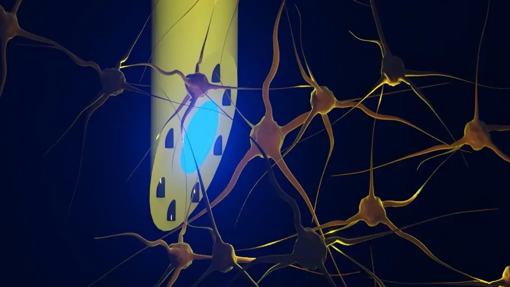
A light-conducting core runs through the center of the fiber, serving as the pathway for optical stimulation. This core is surrounded by eight microscopic channels that are versatile enough to transport liquids—such as targeted medications or chemical tracers—or to house ultra-thin metal wires designed to capture electrical signals from neurons. The resulting fiber measures less than half a millimeter in diameter, making it remarkably compact.
Crucially, the device is highly flexible. Because it is constructed from soft, plastic-like polymers rather than rigid silicon or metal, it possesses a mechanical stiffness that is far closer to that of the brain tissue itself. This allows the implant to move in harmony with the brain’s natural fluctuations, rather than pressing rigidly against delicate tissue. This difference in stiffness is vital, as it significantly mitigates the risk of chronic inflammatory responses that often render harder, more traditional implants ineffective over long periods of implantation.
Tested in Living Mice
The research team successfully validated the system through in vivo experiments conducted on living mice. The electrode was implanted into the rodents’ brains and connected to a comprehensive suite of external equipment, including light sources for optogenetics, recording hardware for electrophysiology, and micro-pumps for the precise delivery of fluids.
The experimental results were striking. The researchers were able to demonstrate that the device could effectively stimulate nerve cells using both blue and red light while simultaneously recording electrical activity from both shallow cortical layers and deeper regions, such as the hippocampus. Furthermore, the team successfully injected different substances at separate depths, with delivery points spaced nearly three millimeters apart. Despite the complexity of these operations, the mice were able to carry the lightweight fiber with no observable signs of discomfort, suggesting the device is well-tolerated even during prolonged testing.
Potential Applications in Epilepsy and Neuroscience
The success of these in vivo trials and the subsequent neurophysiological validation were the result of a deep, collaborative effort. Associate Professor Rune W. Berg from the University of Copenhagen and Associate Professor Rob C. Wykes from University College London provided critical expertise in analyzing neural circuits and developing models relevant to epilepsy. Their involvement ensured that the device’s capabilities were tested against real-world neurological challenges, particularly the complex seizure-generating activity seen in epilepsy models.
The research team is currently in the process of patenting the underlying technology, marking a transition from purely academic exploration to the commercial and clinical development phase. As the group continues to refine the mAxialtrode, they are simultaneously evaluating the regulatory and safety requirements necessary to move the device toward human clinical trials. While the journey from laboratory bench to hospital bedside is arduous, the mAxialtrode represents a significant leap forward in our ability to observe, understand, and eventually modulate the most complex organ in the human body.