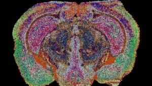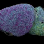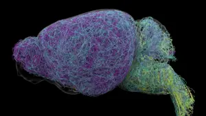








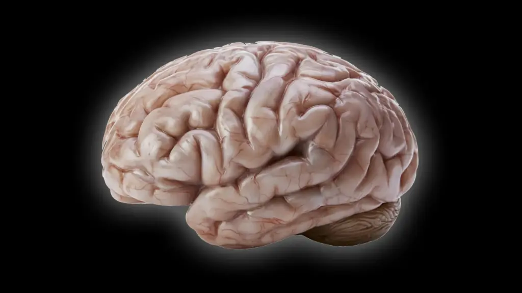
Most primates are characterized by their impressively large, complex brains, which are typically defined by a distinct, rugged landscape of folds and grooves. This intricate architecture, known as gyrification, is a hallmark of mammalian intelligence, providing the surface area necessary to house the billions of neurons that facilitate advanced cognition. Yet, in the diverse family tree of primates, there are notable exceptions. Among them is the common marmoset (Callithrix jacchus), a small, social monkey native to South America whose brain stands in stark contrast to the walnut-like topography of its primate cousins. Its surface is almost entirely smooth—a phenomenon that has long puzzled evolutionary biologists.
Now, researchers at the German Primate Center (DPZ), the Leibniz Institute for Primate Research in Göttingen, have peeled back the layers of this mystery. By peering into the cellular mechanisms that govern early development, the team has identified specific biological processes that explain why the marmoset brain remains smooth while other primate brains evolve into complex, folded structures. The findings, recently published in the journal Science Advances, offer a new window into the evolutionary history of the primate brain and hold significant potential for understanding the developmental disorders that can affect the human brain.
The Evolutionary Puzzle of Brain Folding
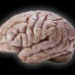
Brain size and the degree of surface folding are not merely aesthetic traits; they are fundamental features that dictate the functional capacity of a species. In humans, the cerebral cortex is heavily folded, a structural adaptation that allows a large amount of neural tissue to fit within the confines of the cranium. These ridges (gyri) and grooves (sulci) act as a biological expansion mechanism, providing space for the extensive connectivity required for complex reasoning, language, and executive function.

For decades, scientists have operated under the assumption that the common ancestor of modern primates possessed a moderately sized brain with at least some degree of folding. However, as primates evolved and diverged into different ecological niches, their brains took on vastly different forms. The common marmoset, which has become a staple of modern biomedical research due to its manageable size and complex social behaviors, presents a fascinating study in evolutionary adaptation. Its brain is nearly lissencephalic, or smooth, a characteristic that requires a departure from the developmental trajectory seen in humans and other large-brained primates.
Understanding why this divergence occurs has been a primary goal for the Junior Research Group Brain Development and Evolution at the DPZ. "We wanted to understand which changes at the cellular level cause the marmoset brain to grow less and form fewer folds," explains Lidiia Tynianskaia, one of the study’s lead authors and a PhD student at the DPZ. "At the beginning of development, the common marmoset brain exhibits the typical structure and composition of a large, folded primate brain. As development progresses, processes must therefore occur that effectively slow down the production of nerve cells."
Uncovering the Cellular Bottlenecks
To pinpoint these processes, the research team employed an innovative methodology using brain organoids—miniature, three-dimensional structures grown from stem cells that mimic the early stages of brain development. By cultivating organoids derived from both human and marmoset cells, the researchers were able to observe the behavior of neural progenitor cells in real-time. These cells are the architects of the brain; they are responsible for generating the neurons that will eventually make up the cerebral cortex. The logic of brain expansion is relatively straightforward: the more neurons these progenitor cells produce, the greater the pressure on the cortex to expand, eventually forcing the tissue to buckle and fold.
The study revealed that the marmoset’s smaller, smoother cerebral cortex is not the result of a single, isolated genetic "switch." Instead, it is the cumulative effect of several distinct developmental differences working in concert. These differences relate to the structure of the progenitor cells, the rate at which they undergo mitosis, and the specific timing of their activity.
"Our investigations have shown that certain progenitor cells in the common marmoset divide significantly more slowly than in humans," notes César Mateo Bastidas Betancourt, the study’s co-lead author and a PhD student at the DPZ. "Other progenitor cells have a simpler structure than their human counterparts, with fewer processes, and are therefore less proliferative. Both of these factors ultimately result in fewer nerve cells, which contributes to a smaller size and less folding of the cerebral cortex in marmosets."
Furthermore, the researchers discovered that the "window of opportunity" for rapid neural growth is significantly curtailed in marmosets compared to humans. In the human developmental timeline, progenitor cells remain active and proliferative for a longer duration, allowing for a massive accumulation of neurons. In marmosets, this period is abbreviated, effectively capping the number of neurons that can be generated before the developmental phase concludes. This combination of slower division rates, simpler cellular architecture, and a shorter growth period provides a comprehensive explanation for the anatomical differences observed in adult marmosets.
The Power of Miniature Models
The use of brain organoids was central to the success of the project. These models provide a unique "in-between" space for researchers, bridging the gap between basic cell culture and the complexities of a living organism. Because studying the fetal development of primates in utero is fraught with ethical and practical challenges—and often produces sample sizes too small for robust statistical analysis—organoids allow researchers to conduct experiments with a higher degree of control and replicability.
Michael Heide, head of the Junior Research Group Brain Development and Evolution, emphasizes the significance of this approach. "The study combines the advantages of in vivo and in vitro methods," says Heide. "Organoids are well-suited for obtaining statistically robust results because such sample sizes are not feasible in primates. We subsequently repeated some key experiments in fetal brain tissue to confirm the results from the organoids."
A critical hurdle in using organoids is ensuring that they accurately reflect the developmental timing of the species from which they are derived. Because the researchers needed to compare human and marmoset development, they had to establish a reliable temporal correlation. Through careful analysis, they determined that a marmoset brain organoid grown for 50 days corresponds to the developmental stage of a natural marmoset brain at 90 days. This calibration was essential, as it allowed the team to interpret their findings with confidence, knowing they were comparing "apples to apples" in terms of biological maturity.
Implications for Evolution and Medicine
The implications of this study extend well beyond the curiosity of primate anatomy. By identifying the specific cellular behaviors that lead to a smooth brain, the researchers have provided a clearer picture of how the wide variety of primate brain shapes emerged over millions of years of evolution. The findings suggest that major evolutionary shifts in brain architecture do not necessarily require the sudden appearance of entirely new genes; rather, they can be achieved through subtle, iterative adjustments to the timing and pace of cellular processes.
Beyond the evolutionary perspective, this research offers a valuable framework for human health. Developmental disorders that affect brain size and structure, such as microcephaly or various cortical malformations, often involve disruptions in the very same progenitor cell populations identified in this study. By mapping the "normal" variations between primate species, scientists are better equipped to identify what goes wrong when these developmental programs are disrupted in humans. Understanding the regulatory mechanisms that control neuron production and brain expansion provides a roadmap for future investigations into the etiology of neurodevelopmental conditions.
As research continues, the common marmoset will likely remain a vital model for understanding the biological constraints and opportunities of the primate brain. Through the marriage of sophisticated organoid technology and comparative developmental biology, the team at the German Primate Center has demonstrated that even the most "simple" brain structures are the product of an incredibly precise and choreographed biological performance—a performance that, when understood, reveals the profound story of how the primate mind came to be.