



The human brain, long considered a relatively static organ once it reaches adulthood, may possess a far more dynamic capacity for self-repair than previously believed. For decades, the prevailing consensus in neurobiology held that once certain foundational support cells within the central nervous system were destroyed—whether through traumatic injury or autoimmune processes—they were effectively lost for good. However, groundbreaking research conducted at the University of Zurich (UZH) is now challenging this rigid paradigm, revealing an extraordinary mechanism by which the adult brain attempts to heal its own damaged architecture.
In a study involving living mouse models, researchers have uncovered that specialized support cells known as astrocytes can orchestrate a sophisticated recovery process. Rather than relying solely on the slow, conventional replication of entire cells, these astrocytes employ an unusual strategy: they produce and dispatch newly formed cell nuclei across significant distances to populate and mend damaged brain regions. This discovery suggests that the brain’s regenerative potential is not only more robust than once thought but also more mechanically complex.
The Critical Role of Glial Cells
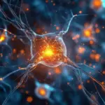
To understand the magnitude of this discovery, it is essential to appreciate the role of glial cells, the unsung workhorses of the nervous system. While neurons often capture the spotlight for their role in transmitting electrical impulses, glial cells—which include the star-shaped astrocytes—are indispensable to the brain’s survival. Astrocytes are named for their characteristic starlike appearance, but their physical structure is matched only by their functional versatility.

These cells serve as the primary caretakers of the brain’s microenvironment. They are deeply involved in maintaining homeostasis, providing essential nutrients to neurons, regulating the intricate flow of blood throughout the brain, and offering structural support to the delicate tissue of the central nervous system. Without healthy, functioning astrocytes, the neural network quickly becomes compromised.
Historically, neuroscientists operated under the assumption that these cells had a limited lifespan and, once decimated, could not be replaced. This perspective has been particularly grim when considering conditions such as traumatic brain injury or autoimmune disorders like neuromyelitis optica spectrum disorder (NMOSD). In NMOSD, the body’s own immune system produces antibodies that specifically target and destroy astrocytes, leading to severe neurological consequences. Because the prevailing view was that the adult brain lacked the plasticity to replace these lost cells, the damage caused by these conditions was frequently viewed as permanent.
Challenging Long-Standing Views on Regeneration
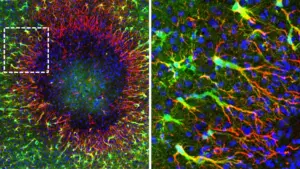
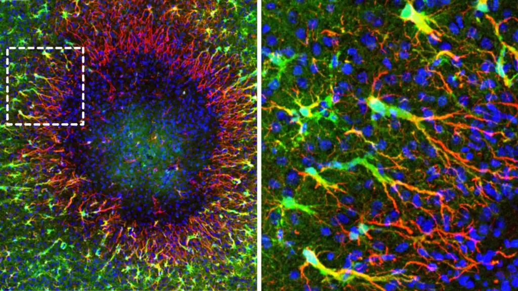
A new study led by co-lead authors Marina Herwerth and Matthias Wyss, under the supervision of Professor Bruno Weber at the Institute of Pharmacology and Toxicology at the University of Zurich, has fundamentally altered this narrative. By observing the brains of living mice, the team identified a specialized population of "regenerative" astrocytes that emerge in the wake of damage.
These regenerative cells appear to congregate strategically around the perimeter of damaged brain tissue. Once positioned at the edges of the injury, they initiate a process of rebuilding the lost astrocyte network. According to Professor Weber, the implications of this discovery are profound. "The findings of our study reveal a previously unknown ability of the adult brain to repair itself," Weber explains. "They point toward new ways of supporting recovery from ailments involving the loss of astrocytes."
The identification of these cells suggests that the adult brain is not merely a passive recipient of injury, but an active participant in its own structural restoration. By characterizing these regenerative astrocytes, the research team has provided a focal point for future medical interventions that aim to harness, rather than merely manage, the brain’s innate healing processes.
A Unique Mechanism: The Migration of Nuclei
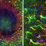
The methodology behind this breakthrough was as innovative as the findings themselves. To track the repair process with the necessary precision, the researchers utilized two-photon microscopy, a sophisticated imaging technique that allowed them to observe the brains of living mice in real time over the course of several weeks. By combining this high-resolution visual tracking with genetic analysis to identify which genes were expressed in specific regions, the team was able to distinguish between standard, stationary astrocytes and the regenerative population responsible for the repair.
What the team observed defied traditional expectations of cell division. Typically, cell regeneration involves a cell dividing into two, with each daughter cell eventually settling into a specific location. In the case of these regenerative astrocytes, however, the process is far more dynamic. The cells undergo a specialized division, after which the newly formed nuclei of the daughter cells literally glide through the long, branching extensions of the astrocyte network.
These nuclei travel considerable distances through the astrocyte scaffold to reach the site of the injury. Once they arrive at the damaged area, they facilitate the repopulation of the lost tissue, effectively knitting the astrocyte network back together. This "gliding" of nuclei represents a previously undocumented dimension of cellular repair, proving that the brain possesses mechanisms for spatial reorganization that scientists had not previously accounted for.
Future Targets for Brain Regeneration
The discovery that cell nuclei can navigate through the complex extensions of adult astrocytes into areas of injury opens a new frontier in regenerative medicine. If scientists can determine the precise conditions under which this process is triggered, they may eventually be able to develop therapeutic strategies to selectively activate these repair mechanisms. Such an advancement could have life-changing implications for patients suffering from conditions characterized by astrocyte loss, offering a path to rebuild neural support networks and improve functional recovery.
Beyond the physical movement of the nuclei, the research team also mapped the genetic landscape of the repair process. They identified a wide array of genes and signaling pathways that become temporarily active only while the repair is underway. These biological signals function as a blueprint for the brain’s recovery efforts, and they represent highly promising targets for pharmacological or gene-based therapies.
"We were able to identify numerous genes and signaling pathways that are temporarily activated during repair," Professor Weber notes. "They could serve as starting points in the future for influencing post-disease and -injury regeneration processes."
By identifying the specific biological cues that initiate and sustain this repair mechanism, researchers hope to create treatments that can "switch on" the brain’s latent regenerative capacity. This would not only provide a more effective way to treat traumatic brain injuries but could also offer a breakthrough in managing chronic autoimmune disorders where astrocyte destruction is a primary driver of disability.
As the scientific community continues to digest these findings, the focus will likely shift toward translating these observations from mouse models to human clinical applications. While the transition from animal research to human medicine is always a complex and cautious process, the discovery of this regenerative pathway provides a concrete foundation for a new era of neuro-regeneration. The brain, it seems, is far more capable of reconstructing itself than we ever dared to imagine, and the "gliding" nuclei of regenerative astrocytes may be the key to unlocking that potential.