







Deep within the brainstem lies a tiny cluster of neurons that exerts a profound influence over the entirety of human experience. Known as the locus coeruleus (LC)—a name derived from the Latin for "blue place" due to its distinct appearance in histological preparations—this region has long been viewed as a monolithic broadcaster. Despite containing only a minuscule fraction of the brain’s total neuronal population, the LC serves as the primary engine for the production of norepinephrine (NE). This vital chemical messenger acts as a neurotransmitter and hormone, modulating everything from our moment-to-moment attention and learning capacity to heart rate, stress responses, and states of excitement.
For decades, neuroscientists operated under a prevailing assumption: the LC functioned much like a high-powered loudspeaker. The working theory held that this small region broadcast a relatively uniform signal across the brain, washing the cortex and other structures in norepinephrine to prepare the organ for arousal or alert states. However, a landmark study published in the journal Nature has fundamentally challenged this "broadcast" model. Through a sophisticated integration of advanced brain imaging, precise neural recording techniques, genetic mapping, and behavioral experiments conducted in mouse models, researchers at the Allen Institute for Brain Science have unveiled that the locus coeruleus is far more complex than previously imagined. Rather than acting as a singular, indiscriminate megaphone, the LC operates as a highly organized, targeted communication network, with distinct neuronal populations tasked with delivering specific signals to localized destinations.
A Hidden Brain Network That Sends Precise Messages
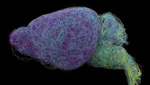
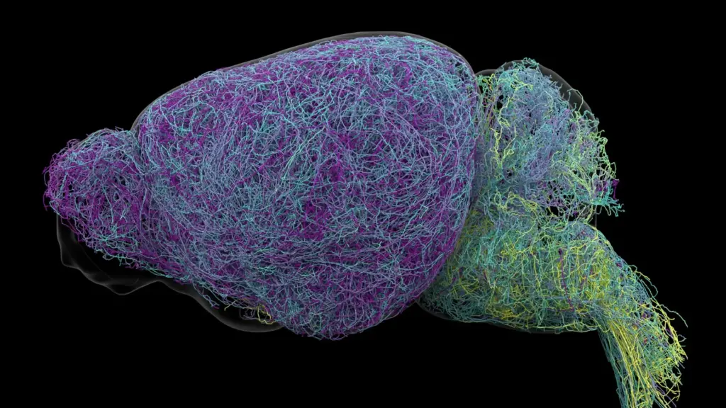
The research team, led by experts at the Allen Institute, employed state-of-the-art tracing techniques to map the axonal projections of LC neurons. Their findings revealed that the LC is not a homogeneous blob of cells, but a structured hub of specialized circuitry. Different groups of neurons within the LC maintain dedicated communication lines to specific regions of the central nervous system.

The architecture of this system follows a clear anatomical division. Neurons located in the upper, or dorsal, portion of the locus coeruleus primarily forge connections with the cerebral cortex and the forebrain—areas of the brain responsible for higher-order cognitive processes. In sharp contrast, cells situated in the lower, or ventral, portion of the structure predominantly project their axons downward toward the brainstem and the spinal cord. This spatial arrangement allows the LC to exert differentiated control over disparate physiological and cognitive functions.
"The findings suggest the brain’s norepinephrine system is far more like a targeted postal network than a foghorn," said Karel Svoboda, director of Neural Dynamics at the Allen Institute and a co-author of the study. By identifying these specific pathways, the researchers have demonstrated that the LC does not simply turn up the volume on brain activity; it directs traffic to ensure that the appropriate signals reach the correct destinations at the right time.
Why the Discovery Could Matter for Brain Disorders
The implications of this discovery extend far beyond basic neuroscience, offering a potential paradigm shift in how clinicians approach various neurological and mental health conditions. Because norepinephrine is central to the regulation of attention, arousal, and mood, it has long been a primary target for pharmacological interventions. Medications used to treat conditions such as attention-deficit/hyperactivity disorder (ADHD), major depressive disorder, and anxiety frequently target the norepinephrine system.
However, current clinical treatments often act as blunt instruments. Because they influence the entire norepinephrine circuit, they frequently produce systemic effects that can lead to unwanted side effects in regions of the brain that do not require modulation. The researchers believe that a clearer, high-resolution map of these specialized LC pathways could eventually pave the way for the development of "circuit-specific" therapies. By selectively targeting the pathways involved in particular symptoms—rather than manipulating the entire norepinephrine system—scientists may be able to design treatments that are both more effective and better tolerated by patients.
Furthermore, the locus coeruleus holds clinical significance in the study of neurodegeneration. LC neurons are among the very first brain cells to exhibit signs of deterioration in Alzheimer’s disease. Given the newfound understanding of the LC’s specialized architecture, this early cell loss may explain why patients experience such a diverse range of cognitive and physiological symptoms. The research, which received substantial support from the National Institutes of Health’s Brain Research Through Advancing Innovative Neurotechnologies (BRAIN) Initiative, provides a foundation for future inquiries into how the progressive breakdown of these specific circuits contributes to the pathology of dementia.
Scientists Identify Brain Cells Involved in Learning
Beyond the anatomical structure, the study uncovered a striking functional division between the dorsal and ventral LC populations. During behavioral tasks, the researchers observed that these two groups responded to different experiences.
The dorsal LC neurons, which are linked to the cortex, showed heightened activity during learning-intensive tasks. Specifically, when mice were engaged in decision-making exercises and were forced to adjust their strategy following an unfavorable or unexpected result, these dorsal neurons surged in activity. This suggests that the dorsal LC is intimately involved in the flexible, adaptive learning process required to navigate changing environments.
The ventral LC neurons, conversely, displayed a distinct behavioral profile. Their activity levels spiked shortly before the mice chose to ignore cues that would normally indicate a potential reward. This suggests that the ventral population acts as a regulator for environmental engagement, helping to determine whether an animal should remain attentive to external stimuli or disengage from its surroundings. "What emerged was a clear map: neurons in the dorsal LC that send signals upward to the cortex are involved in learning, whereas neurons in the ventral region projecting downward to the brainstem and spinal cord govern whether animals engage with their environment at all," Svoboda explained.
The study further demonstrated that these functional differences are reinforced at the genetic level. The researchers found that the anatomical and functional divide is mirrored by distinct gene expression patterns, confirming that the two groups of cells are molecularly distinct and specialized for their respective roles in the brain.
Researchers Discover a Record-Breaking Neuron
To achieve this level of detail, the researchers undertook an exhaustive examination of the mouse brain, utilizing whole-brain imaging to analyze nearly 35,000 neurons and profiling the genetic signatures of almost 400,000 individual cells. Among these reconstructions, they identified an extraordinary physical characteristic of LC neurons: the possession of exceptionally long axons.
Axons are the slender fibers that allow neurons to carry electrical signals across long distances. While many neurons in the brain have relatively localized reach, the researchers found that LC axons measured, on average, 35 centimeters—a staggering distance for a structure as compact as a mouse brain. The team reconstructed one particularly remarkable neuron that reached a total length of 70.32 centimeters, marking it as one of the longest single neurons ever documented in a mouse model.
"This neuron, like many others that we studied, supplies norepinephrine to a very large volume of the cerebral cortex," said Jeremiah Cohen, an Allen Institute scientist and co-author of the study. "For the brain, this is highly unusual. Most neurons are more specific in their targets." Despite this massive reach, however, the neuron remains highly selective. The researchers noted that this specific cell purposefully avoids releasing norepinephrine in the cerebellum, brainstem, or spinal cord, further reinforcing the idea that even these wide-reaching neurons follow a highly organized, targeted distribution plan.
A New Understanding of How the Brain Learns
The study also draws intriguing parallels between the norepinephrine system and the brain’s dopamine signaling pathways. Dopamine is widely recognized for its role in the basal ganglia, a region of the brain critical for the formation of habits and the reinforcement of behavior. The researchers hypothesize that norepinephrine and dopamine may operate in tandem, forming a collaborative network that allows the brain to acquire and integrate complex, abstract concepts simultaneously.
By mapping the intricate connections, functional specialization, and the unusual physical architecture of the locus coeruleus, this research provides a significantly more nuanced picture of how the mammalian brain communicates, learns, and regulates its response to the world. While these insights were gained through experiments in mice, the fundamental organization of these circuits offers a new roadmap for researchers to investigate human brain function. As the field moves forward, the hope remains that these detailed anatomical and functional insights will translate into more precise, targeted clinical interventions for the millions of people living with neurological and psychiatric disorders.