







In the intricate landscape of primate evolution, the architecture of the brain stands as a testament to the diverse paths life can take. While most primates are characterized by relatively large, complex brains adorned with deep folds and grooves—a hallmark of cognitive sophistication—the common marmoset (Callithrix jacchus) serves as a striking evolutionary outlier. These small South American monkeys possess brains that are remarkably smooth, lacking the extensive folding patterns that define the human cerebral cortex. Now, researchers at the German Primate Center (DPZ), the Leibniz Institute for Primate Research in Göttingen, have unveiled the cellular mechanisms that account for this profound difference, offering a new window into the evolutionary history of the primate brain.
The research, recently published in the journal Science Advances, illuminates how subtle variations in the behavior of neural progenitor cells dictate the final shape and size of the brain. By peering into the microscopic world of brain organoids, the scientific team has identified specific cellular processes—involving structure, division speed, and developmental timing—that ultimately limit the number of neurons produced in marmosets, thereby resulting in their smaller, smoother cortical surfaces.
Why Some Primate Brains Are Smooth Instead of Folded
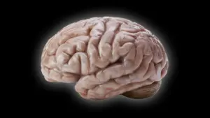
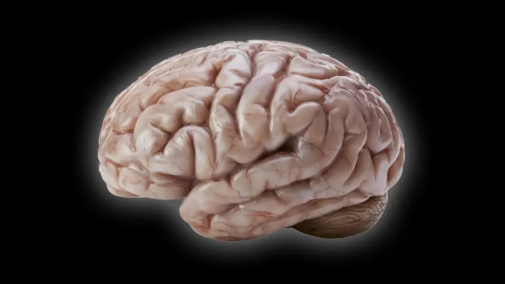
To understand the magnitude of this discovery, one must consider the functional anatomy of the brain. In primates like humans, the cerebral cortex is highly folded, resembling the intricate ridges and valleys of a walnut. This folding, known as gyrification, is not merely a structural quirk; it is a critical biological strategy. By folding the surface of the brain, nature expands the available surface area within the confined space of the cranium, allowing for the packing of billions of additional neurons. These neurons are the engine of complex cognition, facilitating advanced mental processes, problem-solving, and social behavior.

For years, evolutionary biologists have operated under the assumption that the common ancestor of modern primates possessed a brain of moderate size that already exhibited at least some degree of folding. However, the lineage leading to the common marmoset took a different trajectory. As these monkeys became a cornerstone of modern biomedical research, the need to understand their neurodevelopmental profile grew. Researchers at the German Primate Center set out to bridge the gap in knowledge regarding why the marmoset brain diverges so sharply from the human model.
"We wanted to understand which changes at the cellular level cause the marmoset brain to grow less and form fewer folds," explains Lidiia Tynianskaia, one of the study’s two first authors and a PhD student in the Junior Research Group Brain Development and Evolution at the DPZ. "At the beginning of development, the common marmoset brain exhibits the typical structure and composition of a large, folded primate brain. As development progresses, processes must therefore occur that effectively slow down the production of nerve cells."
Scientists Discover What Slows Brain Growth
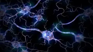
To investigate these developmental nuances, the research team employed an innovative methodology, utilizing brain organoids grown from human and common marmoset cells. These "mini-brains" are three-dimensional structures that mimic the developmental architecture of the early brain, providing a controlled environment to study the life cycle of neural progenitor cells. These progenitor cells are the architects of the brain; they are responsible for generating the neurons that will eventually form the cerebral cortex. The logic of neurodevelopment is straightforward yet precise: the more neurons these progenitor cells produce, the greater the potential for the cortex to expand and fold as the brain grows.
The study revealed that the marmoset’s relatively smooth cortex is not the result of a single genetic "switch," but rather a culmination of several distinct cellular differences. These differences work in tandem to curtail the production of nerve cells.
"Our investigations have shown that certain progenitor cells in the common marmoset divide significantly more slowly than in humans," notes César Mateo Bastidas Betancourt, the study’s co-first author and a PhD student in the Junior Research Group. "Other progenitor cells have a simpler structure than their human counterparts, with fewer processes, and are therefore less proliferative. Both of these factors ultimately result in fewer nerve cells, which contributes to a smaller size and less folding of the cerebral cortex in marmosets."
Beyond the speed of division and the morphology of the cells, the timing of development emerged as a crucial factor. The researchers identified significant differences in the temporal window during which these progenitor cells remain active. Compared to humans, marmosets have a much shorter period of rapid cell proliferation. When the "window" for producing neurons closes prematurely, the brain simply lacks the raw material—the neurons themselves—required to drive the expansion and folding processes that characterize larger, more complex brains.
Miniature Brains Reveal Important Developmental Clues
The reliance on brain organoids was fundamental to the success of the study. Growing these organoids allowed the researchers to conduct robust, statistically significant comparisons that would be impossible to achieve through fetal tissue analysis alone. By scaling their observations across multiple samples, the team could isolate specific variables in cell behavior with a high degree of precision.
However, the team did not rely solely on these laboratory models. To ensure their findings were not artifacts of the organoid environment, the researchers validated their results by replicating key experiments in actual fetal brain tissue. This dual approach—combining the advantages of in vitro models with in vivo confirmation—provided a level of rigor rarely seen in such comparative studies.
A pivotal moment in the research involved establishing a "developmental clock" between the organoids and the actual organisms. The researchers discovered that a marmoset brain organoid cultured for 50 days mirrors the developmental stage of a natural marmoset brain at 90 days. Establishing this chronological relationship was essential; it allowed the scientists to translate their findings from the petri dish to real-world biology, ensuring that the developmental milestones they were observing were accurate and comparable across both systems.
"The study combines the advantages of in vivo and in vitro methods," says Michael Heide, head of the Junior Research Group Brain Development and Evolution. "Organoids are well-suited for obtaining statistically robust results because such sample sizes are not feasible in primates. We subsequently repeated some key experiments in fetal brain tissue to confirm the results from the organoids."
What Smooth Monkey Brains Reveal About Human Brain Evolution
The implications of this research extend far beyond the anatomy of the marmoset. By identifying how small, granular changes in cell behavior—such as the speed of a division cycle or the timing of a developmental phase—can lead to massive, macroscopic differences in brain architecture, the study provides a roadmap for understanding the evolution of the primate brain. It suggests that the diversity of brain shapes observed across the primate order is not necessarily the result of creating "new" types of cells, but rather the result of "tuning" the existing developmental program.
For evolutionary biologists, this is a significant step forward in explaining why some species evolved toward cognitive complexity through brain expansion and folding, while others thrived with more compact, smooth-brained architectures.
Furthermore, the study holds promise for the field of human medicine. By mapping the cellular mechanics that control neuron production and brain growth, the research establishes a foundational understanding of normal development. This knowledge is indispensable for future investigations into neurodevelopmental disorders in humans, where these very processes may go awry. If researchers can pinpoint the cellular mechanisms that govern the healthy growth of the cortex, they may eventually be able to better understand the origins of conditions where brain development is disrupted or altered.
As the scientific community continues to explore the complexities of the brain, the common marmoset—once merely a model organism—has provided a vital piece of the evolutionary puzzle. The humble, smooth brain of the marmoset, when held against the mirror of the human brain, reflects the profound power of developmental timing and cellular rhythm in shaping the biological world. Through the lens of the German Primate Center’s latest findings, we are one step closer to understanding the mechanisms that built the most complex structure in the known universe.