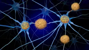









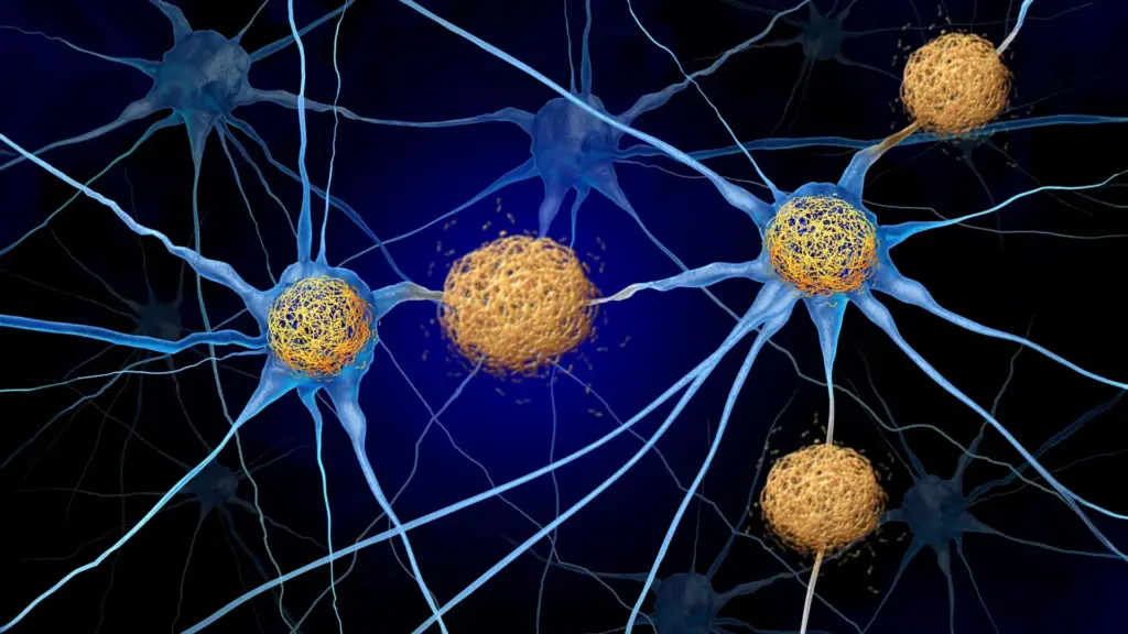
Protecting brain cells from the progressive destruction that characterizes Alzheimer’s disease remains one of the most formidable challenges in modern medicine. For decades, the primary focus of neurodegenerative research has been the physical buildup of proteins—specifically amyloid plaques and tau tangles—that accumulate in the brains of affected individuals. These disorders, collectively known as tauopathies, are marked by the formation of abnormal tau protein structures inside neurons. As these tangles proliferate, nearby brain cells begin to wither and die, leading to the devastating loss of memory and independence that defines the condition.
However, a growing body of evidence is beginning to challenge the long-held assumption that tau protein accumulation is the sole driver of neuronal death. Scientists are increasingly looking toward the brain’s own neighborhood—the immune system—to explain the rapid loss of cognitive function. New findings suggest that it is not the tau tangles themselves that are responsible for the bulk of the cellular destruction, but rather the brain’s inflammatory response to these proteins. By identifying a specific pathway that guides immune cells into the brain, researchers at the Washington University School of Medicine in St. Louis have uncovered a potential new therapeutic strategy that could transform the treatment landscape for Alzheimer’s and related conditions.
A Breakthrough in Interrupting the Immune Response
The research, published September 28 in the journal Neuron, highlights a promising method for intercepting the immune system’s harmful reaction to tau accumulation. Led by a team at WashU Medicine, the study utilized a mouse model designed to mimic the tau buildup seen in human Alzheimer’s patients. The researchers focused their attention on a protein called CXCR3, which acts as a molecular "beacon" or receptor on the surface of T cells—a type of immune cell—that allows them to navigate into the brain.

By administering an antibody designed to block the CXCR3 receptor, the researchers effectively severed the communication line between the brain’s distress signals and the invading immune cells. The results were dramatic. Over the course of several months, the number of T cells that successfully infiltrated the brain in treated mice was reduced by approximately 50% compared to a control group.
The clinical outcomes were even more significant. Despite the fact that the actual volume of accumulated tau in the brains remained unchanged, the mice receiving the antibody showed a 40% higher retention of brain tissue in regions critical for memory. Furthermore, these mice demonstrated superior performance on cognitive tests compared to their untreated counterparts. This finding suggests that even when the underlying pathology—the tau tangles—persists, the associated neurodegeneration can be significantly mitigated by dampening the immune system’s misguided attack.
"In tauopathies, including Alzheimer’s disease, there’s no treatment right now that actually decreases neurodegeneration," explained David M. Holtzman, MD, the senior author of the study and the Barbara Burton and Reuben M. Morriss III Distinguished Professor in the Department of Neurology at WashU Medicine. "If we can show that we’re really decreasing brain cell death, it’s certainly worth trying to pursue that pathway to prevent the most devastating consequences of these diseases."
Reevaluating the Immune System’s Role in Alzheimer’s
To understand the gravity of these findings, it is necessary to examine the current standard of care for Alzheimer’s disease. Today’s most advanced therapies, such as the FDA-approved medications lecanemab and donanemab, are designed to clear amyloid plaques—deposits that form between neurons during the early stages of the disease. These plaques are known to interfere with synaptic communication and are thought to contribute to the subsequent formation of tau tangles.
While these drugs have shown success in slowing cognitive decline, they have not yet demonstrated an ability to fully arrest the death of neurons. Moreover, these treatments are largely ineffective for primary tauopathies—diseases where tau buildup occurs independently of amyloid deposits. Because Alzheimer’s involves both proteins, it is categorized as a secondary tauopathy, yet the persistent neuronal loss suggests that clearing amyloid is only part of the puzzle.
Dr. Holtzman’s laboratory has spent years investigating whether the immune system might hold the key to addressing the gaps in current treatment. A 2023 study published in Nature by the same group revealed that T cells do not merely wander into the brain at random; they specifically concentrate in regions heavily populated with tau tangles, acting as active participants in the neurodegenerative process. A subsequent study published earlier this month in Nature Neuroscience further refined this understanding, identifying that the chemical signals directing these T cells are actually generated in the lymph nodes located outside the brain, rather than within the brain itself.
Deciphering the Pathway: The CXCL10-CXCR3 Axis
With the knowledge that immune cells were contributing to the damage, the researchers sought to understand the "GPS" system used by T cells to infiltrate the brain. T cells navigate the body by following chemical signals known as chemokines. Previous research in the Holtzman lab had identified an abundance of one such chemokine, CXCL10, in the brains of mice with tau accumulation—a finding mirrored in human patients with Alzheimer’s.
The relationship is clear: activated T cells carry the CXCR3 receptor, which acts like a lock that fits perfectly with the CXCL10 key. This CXCL10-CXCR3 pathway creates a chemical trail that leads harmful T cells directly into the brain tissue. By studying mice lacking either the CXCL10 signal or the CXCR3 receptor, the researchers confirmed that T cells were unable to infiltrate the brain, even when the animals were subjected to inflammation that would normally trigger an immune response. This confirmation provided the necessary evidence to move toward an experimental treatment.
Therapeutic Implications: Treating From the Outside In
The success of the antibody treatment in preserving brain tissue and memory function opens a new frontier in drug development. Perhaps most significantly, the researchers noted that the antibody reached the boundary of the brain but did not need to cross the blood-brain barrier to be effective.
The blood-brain barrier is a highly selective semipermeable border that protects the brain from pathogens but also prevents the vast majority of therapeutic drugs from reaching their targets. If a treatment can be effective by targeting immune cells from outside the brain—or by blocking the signals that originate in the lymph nodes—the challenges associated with drug delivery are drastically reduced.
"For this therapeutic approach, if it is safe, you wouldn’t have to design the drug to get into the brain—which is a big deal since most molecules don’t cross the blood-brain barrier well—and you don’t have to get rid of the tau to get this therapeutic effect," Dr. Holtzman noted.
A New Horizon for Alzheimer’s Care
While these results in animal models are undoubtedly encouraging, the research team is cautious about immediate clinical application. Additional rigorous testing is required to ensure that such an approach is safe for humans. However, the prospect of repurposing existing medications offers a potential shortcut. There are already several drugs used to manage T-cell activity in patients with autoimmune disorders like multiple sclerosis. Researchers believe these existing treatments could serve as a vital starting point for clinical trials aimed at reducing neurodegeneration in Alzheimer’s patients.
This shift in strategy represents a fundamental change in how the medical community views neurodegenerative diseases. By moving beyond a singular focus on protein clearance and beginning to address the secondary inflammatory damage caused by the immune system, scientists are opening doors to more comprehensive treatment plans.
"Tauopathies aren’t thought of as autoimmune disorders, so they haven’t been treated the same way, but this study shows for the first time in an animal model that these diseases respond to a specific T-cell therapy," said Dr. Holtzman, who also serves as the director of the Knight Alzheimer Disease Research Center and the Hope Center for Neurological Disorders at WashU Medicine.
As the scientific community continues to explore the complex relationship between the brain and the immune system, the findings from this study provide a glimmer of hope. By targeting the immune response, clinicians may eventually be able to preserve the cognitive integrity of patients, even in the presence of the protein tangles that have long served as the primary target of Alzheimer’s research. This research, supported by various organizations including the National Institutes of Health, the BrightFocus Foundation, and private philanthropic initiatives, marks a significant step toward a future where Alzheimer’s and other tauopathies may one day be managed as chronic, treatable conditions rather than inevitable, progressive losses of the self.