






You walk out of the grocery store, the cool evening air hitting your face as you prepare for the commute home. You reach for your keys, your mind already turning toward dinner, when you stop abruptly in the middle of the parking lot. You scan the rows of vehicles, but the mental image you need—the specific spot where you parked just twenty minutes ago—simply isn’t there. It is a common, frustrating experience that most of us dismiss as a momentary lapse in focus. Yet, for many, this type of memory lapse is a symptom of a deeper, more complex biological process unfolding within the brain.
Our ability to recall personal experiences, a function known as episodic memory, is the bedrock of our daily lives. From the emotional weight of a childhood milestone, like the first day of elementary school, to the mundane task of remembering where you parked your car, these memories rely on the brain’s capacity to encode and store information. As we age, however, this capacity often begins to wane. The intricate web of sights, sounds, and contextual details that comprise our experiences becomes increasingly difficult to retrieve. While scientists have long recognized that some degree of cognitive decline is a standard part of aging, the degradation can become far more severe in individuals developing Alzheimer’s disease.
A groundbreaking study led by researchers at the University of California, Berkeley, is now shedding new light on the biological mechanisms behind this decline. By investigating the architecture of the aging brain, neuroscientist Omer Sharon and his team, working under the guidance of renowned sleep researcher Matthew Walker, have uncovered a critical connection between disrupted brain waves during deep sleep, the accumulation of the Alzheimer’s-associated protein tau, and a marked decline in memory performance.
How Deep Sleep Orchestrates Memory Consolidation
Sleep is not a uniform state; it is a complex landscape of distinct stages, each defined by unique patterns of electrical activity. While rapid eye movement (REM) sleep is often associated with vivid dreaming and eye movement, non-REM sleep—particularly its deeper stages—is vital for maintaining cognitive health. It is during these periods of deep, dream-limited rest that the brain performs essential "housekeeping" tasks, including the consolidation of memories.
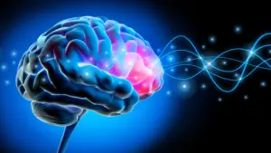
During deep sleep, the brain undergoes a process of profound synchronization. Large populations of neurons switch off and back on in a highly coordinated, rhythmic sequence. These coordinated shifts generate slow electrical waves that typically originate in the frontal cortex and ripple across the surface of the brain.
"Each slow wave reflects an enormous population of neurons switching off and back on together," explains Sharon, a postdoctoral researcher at UC Berkeley’s Center for Human Sleep Science. "Our study shows that for memory, it’s critical that these events cascade in sequence across large parts of the brain. This coordinated shutdown has been observed only during deep sleep, and may be one reason why we sleep."
The study, published in the journal Nature Neuroscience, suggests that the accumulation of tau—a protein linked to the pathology of Alzheimer’s—disrupts this delicate synchronization. As tau builds up in the frontal cortex, these sweeping waves of electrical activity lose their cohesion. Instead of traveling in synchronized, broad patterns, the waves become scattered and restricted in distance. This fragmentation, the researchers argue, is a primary driver of weakened memory consolidation and declining cognitive function.
The Link Between Tau and Disrupted Electrical Activity
To test this hypothesis, the UC Berkeley team conducted a rigorous comparison of brain activity in two distinct groups: cognitively healthy young adults in their early 20s and older participants between the ages of 65 and 75. Using electroencephalograms (EEGs) to monitor sleep, the researchers observed a stark contrast in neurological behavior.
In the younger cohort, slow waves moved in powerful, coordinated groups across distances equivalent to the width of a human hand on the scalp. In contrast, the older adults exhibited waves that traveled significantly shorter distances and appeared to fire independently, lacking the rhythmic harmony seen in the youth.
To understand the role of tau, the researchers collaborated with Dr. William Jagust, a professor emeritus of public health and neuroscience at UC Berkeley. Using positron emission tomography (PET) scans, which employ radioactive tracers to visualize biological substances within the brain, the team mapped the distribution of tau in the older participants. The data revealed a clear correlation: higher levels of tau in the frontal cortex directly corresponded to a breakdown in the longer, traveling waves observed during deep sleep.
Crucially, these findings were observed in participants who had not yet been diagnosed with Alzheimer’s disease. "These people did not have Alzheimer’s," Sharon noted. "They had tau in their brains, but with subclinical impacts. Their memory decline was within the normal range for their age." This suggests that the physiological precursors to memory loss begin long before clinical symptoms manifest, with tau accumulation serving as a silent indicator of future cognitive struggle.
The Emergence of "Lonely Waves"
The team sought to determine if these physiological disruptions had a tangible impact on the participants’ ability to retain information. In a series of memory tests, participants were taught pairs of associated words in the evening. The following day, researchers tested the participants’ recall to measure how effectively the information had been consolidated overnight.
The results were telling. Participants who displayed shorter, more isolated slow waves—what the researchers dubbed "lonely waves"—consistently struggled to recall the word associations. The correlation between wave pattern and memory retention was significant, suggesting that when the brain’s "memory-storing" electrical activity becomes fragmented, the ability to lock in new information overnight is severely compromised.
Furthermore, by tracking a subset of the participants over several years, the researchers were able to observe the progression of these changes. As frontal cortex tau levels increased over time, the slow waves became progressively less coordinated, and the participants’ ability to retain new information experienced a corresponding decline. While these findings provide strong evidence of a link between tau, sleep, and memory loss, the researchers emphasize that they do not yet prove a direct causal relationship. It remains to be seen whether tau causes the waves to break down, or if the disruption of these waves facilitates the accumulation of tau.
Strengthening the Evidence Through Spinal Fluid Analysis
Given the high cost and limited accessibility of PET imaging, the researchers sought to validate their findings using a different biological marker. Partnering with neurologist Yo-El Ju at Washington University in St. Louis, the team analyzed spinal fluid from a separate cohort of older adults.
While spinal fluid analysis cannot pinpoint the specific location of tau accumulation in the frontal cortex, it provides an accurate measure of Alzheimer’s-related proteins throughout the central nervous system. The results confirmed the initial findings: individuals whose sleeping brains produced more "lonely waves" showed higher ratios of tau to amyloid—another protein characteristic of Alzheimer’s—in their spinal fluid.
"It’s collected in a totally different place and in a different cohort, so I think that adds to the strength of the evidence," Sharon said. The consistency across independent groups and distinct testing methodologies underscores the validity of the link between tau pathology and the disruption of deep-sleep electrical patterns.
Future Implications for Brain Health
The findings from the UC Berkeley study suggest that the cognitive decline associated with aging is not merely an inevitable consequence of time, but a process heavily influenced by specific pathological changes. By identifying that tau buildup in the frontal cortex acts as a catalyst for the breakdown of global sleep waves, researchers have identified a potential pathway for intervention.
"It’s not just about age," Sharon remarked. "It’s how much pathology you have in frontal areas where global waves originate."
As researchers look toward the future, the ability to monitor and potentially stabilize these slow-wave patterns could offer new ways to identify early signs of memory impairment. While the fundamental question of whether the "lonely waves" are a symptom or a cause remains to be answered, the study provides a vital roadmap for future research. Understanding how these biological markers interact will be essential for designing treatments aimed at protecting the aging brain and preserving the integrity of memory.
The research team, which included co-authors Xi Chen, James Westphal, Chelsea Brown, and Vyoma D. Shah of UC Berkeley, along with Jason Dude of Washington University, was supported by funding from the National Institute on Aging at the National Institutes of Health, with additional fellowships provided by the Glenn Foundation for Medical Research and the Weill Neurohub. As the scientific community continues to explore the mysteries of the sleeping brain, this work stands as a testament to the complex, silent, and vital work the brain performs while we rest.