






Long before a human being draws their first breath, the brain is assembled through a staggeringly complex series of cellular decisions. At the very heart of this architectural marvel are radial glia—a specialized class of stem cells that serve as the master builders of the human cerebral cortex. These cells are responsible for generating the vast majority of neurons and support cells that define the region of the brain associated with complex thought, memory, and language.
The role of radial glia extends beyond simple construction; they are believed to be the primary drivers of the extraordinary expansion of the human cortex compared to that of other species. While these cells largely vanish before birth, they retain a mysterious connection to our health throughout our lives. For reasons that continue to elude researchers, similar cellular programs can re-emerge in brain cancers, suggesting that the "instructions" these cells use to build a brain may also be hijacked to fuel malignant growth.
"Radial glia are the coolest cells that have ever existed," says Aparna Bhaduri, an assistant professor of biological chemistry at the David Geffen School of Medicine at UCLA. "They’re really key to making us human. But they’re also at the center of many neurodevelopmental and neuropsychiatric disorders, as well as cancer—so understanding how they make their decisions is one way to start understanding how those conditions arise."
Two landmark studies, recently published in the journals Cell and Science, have provided an unprecedented look into the decision-making processes of these foundational cells. By examining how radial glia respond to metabolic shifts and direct physical contact from other regions of the developing brain, Bhaduri and her team have uncovered new insights into the cellular variety that characterizes the human cortex.
Metabolism as a Master Regulator of Brain Development
In the study published in Cell, researchers sought to move beyond the traditional view of metabolism as a passive provider of energy. Led by co-first authors Jessenya Mil and Jose Soto, in collaboration with the laboratory of Heather Christofk, the team constructed a comprehensive, high-resolution map of metabolism within the developing human cortex.
To build this metabolic atlas, the researchers analyzed donated human tissue alongside brain organoids—miniature, three-dimensional models of brain tissue grown from stem cells in the laboratory. The results were striking: metabolism does not merely fuel the background operations of a cell; it actively dictates the cell’s developmental trajectory.
The study revealed that radial glia rely heavily on the pentose phosphate pathway, a metabolic process that redirects glucose to produce the essential building blocks required for rapidly dividing cells. When the researchers experimentally lowered glucose availability or disrupted this specific metabolic pathway, the stem cells underwent a behavioral shift. They moved away from their typical production patterns and began generating inhibitory neurons and other cell types that, under normal conditions, would only appear much later in the developmental timeline.
"What was surprising is that metabolism isn’t just a passive thing that happens in the background," explains Bhaduri, who is a member of both the UCLA Broad Stem Cell Research Center and the UCLA Health Jonsson Comprehensive Cancer Center. "It can really control how stem cells make decisions."
This discovery has profound implications for understanding how environmental factors, such as maternal nutrition or metabolic disorders, might influence fetal brain development. Furthermore, the metabolic atlas serves as a critical resource for the scientific community, offering one of the most granular maps to date of how energy processing evolves alongside human neural maturation.
Thalamic Signals and the Architecture of the Cortex
While the first study focused on the internal chemistry of the cell, the second study—published in Science and led by first author Claudia Nguyen—looked outward. The researchers investigated the role of external signals originating from the thalamus, a central structure deep within the brain that serves as a vital relay station for the nervous system.
For years, neuroscientists have understood that thalamic neurons extend long, wire-like projections toward the cortex, eventually forming precise connections. However, human anatomical studies have long noted a peculiar timeline: these fibers arrive at the cortex long before the actual synaptic connections are fully formed. This raised a persistent question in the field: why do these projections arrive so early?
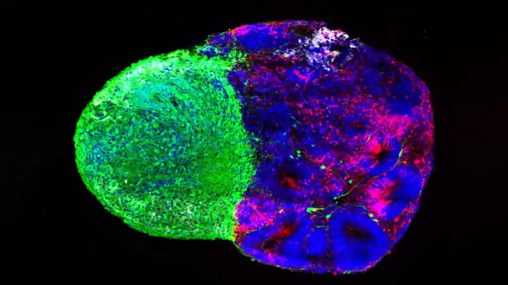
Using sophisticated human stem cell-derived "assembloids"—complex models that combine different types of brain tissue to mimic how regions interact—the UCLA team discovered that the thalamic projections physically touch radial glia while the brain is still in its infancy. This physical interaction is a transformative event for the stem cells. Upon contact, the radial glia are prompted to produce more excitatory neurons, which serve as the primary signal-carrying cells in the cortex. This effect was particularly pronounced in the upper layers of the cortex, an area that has undergone significant expansion during human evolution.
"We already knew that these projections influence how the cortex develops," Bhaduri notes. "What we specifically found is that this influence comes through an actual physical connection between the projections and the radial glia—a point of contact that just hasn’t been identified before, and one that very likely does not exist in rodents."
Genetic Vulnerability and the Spectrum of Development
The importance of this physical signaling was further underscored by investigating the NRXN1 gene. Already well-established in the scientific literature for its role in helping neurons form connections, mutations in NRXN1 are known to be linked to autism spectrum disorder.
When the team created assembloids using patient-derived cells carrying an NRXN1 mutation, they observed a significant deviation from normal development. The thalamic signals, altered by the genetic deficiency, triggered a different response in the radial glia. This caused a shift in the balance between the pool of available stem cells and the resulting neurons they generated, suggesting a mechanism by which early developmental disturbances can cascade into complex neurodevelopmental conditions.
A New Era for Organoid Research
The two studies, while distinct in their approach, converge on a fundamental truth: radial glia are not solitary actors. They operate within a constant, dynamic dialogue with their environment, whether through the metabolic fuel they consume or the physical touch of distant neural projections.
The research also highlights the rapid evolution of organoid technology. Only a decade ago, the ability to observe these nuanced human developmental processes in real-time was virtually non-existent. Today, brain organoids and assembloids have democratized the field, allowing scientists to ask questions that were once confined to the realm of theoretical models or limited by the constraints of animal research.
Bhaduri hopes these findings will shift the paradigm in developmental biology, encouraging her colleagues to view metabolism and cellular contact as active drivers of development rather than mere background noise. By pulling back the curtain on how these decisions are made at the cellular level, researchers are taking the first steps toward a more profound understanding of the human brain—from its normal, healthy development to the origins of disease vulnerability and the mysterious, aggressive behavior of cancer stem cells.
The research was supported by a wide range of organizations, including the National Institutes of Health, the National Science Foundation, the Brain & Behavior Research Foundation, the Alfred P. Sloan Foundation, the Rose Hills Foundation, the Esther A. & Joseph Klingenstein Fund, the Simons Foundation, the Chan Zuckerberg Initiative, the NIH BRAIN Initiative Cell Atlas Network, the International Foundation for Ethical Research, the UCLA Broad Stem Cell Research Center’s Stem Cell Research Training Program, and the UCLA Health Jonsson Comprehensive Cancer Center and UCLA Broad Stem Cell Research Center Ablon Scholars Program.