







Long before a human infant takes their first breath, their brain undergoes an architectural transformation of staggering complexity. This prenatal assembly process is defined by an enormous series of cellular decisions, a sequence of events that dictates the structure and function of the human mind. At the very center of this biological masterclass are radial glia—a specialized class of stem cells that serve as the primary architects of the human brain, responsible for creating the unique features that distinguish our species from all others.
These remarkable cells are the progenitors of the cerebral cortex, the sophisticated, folded region of the brain that governs higher-order functions including complex thought, memory, and language. Radial glia are widely believed to be the driving force behind the dramatic expansion of the human cortex compared to the brains of other mammals. While these cells are primarily active during fetal development and largely disappear before birth, their legacy persists in unexpected ways. Scientists have observed that similar cellular mechanisms can re-emerge in brain cancers, a phenomenon that remains a profound mystery in modern oncology.
"Radial glia are the coolest cells that have ever existed," says Aparna Bhaduri, an assistant professor of biological chemistry at the David Geffen School of Medicine at UCLA. "They’re really key to making us human. But they’re also at the center of many neurodevelopmental and neuropsychiatric disorders, as well as cancer—so understanding how they make their decisions is one way to start understanding how those conditions arise."
To bridge the gap between abstract biological potential and the physical reality of the human brain, researchers have long sought to uncover the specific "instructions" these cells follow. Now, two groundbreaking studies published in the journals Cell and Science have provided an unprecedented look into how radial glia navigate their developmental paths. By investigating how these cells respond to both internal metabolic states and external physical signals, Dr. Bhaduri and her colleagues have unveiled new insights into the mechanisms that produce the vast, intricate variety of cell types found within the human cortex.
Metabolism as a Directive Force in Brain Development
In the study published in Cell, researchers set out to map the metabolic landscape of the developing human cortex. This ambitious project, a collaboration between the labs of Dr. Bhaduri and Heather Christofk, and led by co-first authors Jessenya Mil and Jose Soto, sought to determine whether metabolism acts merely as a passive fuel source or as an active controller of cellular fate.
To create this comprehensive metabolic atlas, the team analyzed donated human tissue alongside brain organoids—miniature, three-dimensional models of the brain grown in the laboratory from human stem cells. The results challenged conventional wisdom: metabolism does not simply provide the energy required for brain development to occur in the background; it actively influences the identity and behavior of the cells being produced.
The researchers discovered that radial glia rely heavily on a metabolic process known as the pentose phosphate pathway. This pathway utilizes glucose to synthesize the essential building blocks required by cells that are in a state of rapid division. When the scientists experimentally limited the availability of glucose or disrupted the pentose phosphate pathway, the radial glia underwent a significant shift in their developmental output. Rather than continuing their standard production cycle, the stem cells began generating different cell types, specifically inhibitory neurons and other varieties that typically appear much later in the developmental timeline.
"What was surprising is that metabolism isn’t just a passive thing that happens in the background," says Bhaduri, who is a member of both the UCLA Broad Stem Cell Research Center and the UCLA Health Jonsson Comprehensive Cancer Center. "It can really control how stem cells make decisions."
The implications of this finding are far-reaching. By identifying metabolism as a regulatory switch, scientists now have a new framework to investigate how maternal nutrition, metabolic disorders, and various environmental factors might alter the trajectory of fetal brain development. Furthermore, this new metabolic atlas serves as a vital resource for the global research community, offering a high-resolution look at the energetic requirements of the human brain as it builds itself from the ground up.
The Influence of Physical Signals from the Thalamus
While the metabolic study examined the internal "fueling" of development, the second study, published in Science and led by first author Claudia Nguyen, explored a completely different source of information: physical contact. The researchers focused their attention on the thalamus, a critical structure located deep within the brain that functions as a central relay station, transmitting sensory and motor information throughout the nervous system.
For decades, neuroscientists have known that neurons within the thalamus extend long, wire-like fibers, known as axons, toward the cortex. Eventually, these fibers form precise connections with specific cortical neurons. However, previous anatomical studies revealed a curious timing mismatch: in humans, these thalamic projections arrive at the cortex long before the final synaptic connections are actually established. This raised a fundamental question in developmental biology: Why do these fibers arrive so early, and what are they doing while they wait to connect?
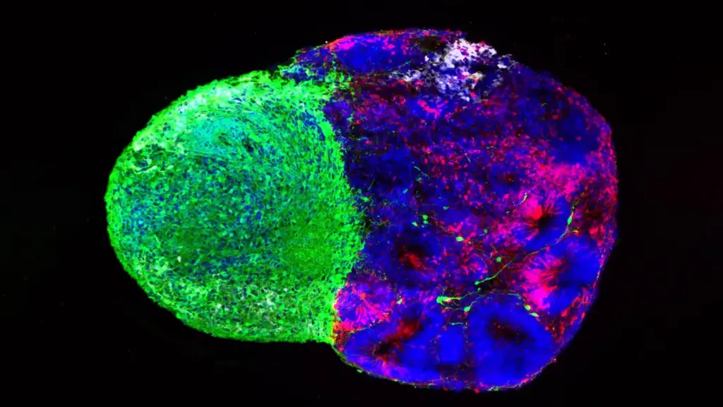
Using advanced human stem cell-derived "assembloids"—complex models where different parts of the brain are grown together to mimic their natural interactions—the UCLA team found a compelling answer. They observed that the thalamic projections physically touch the radial glia while the brain is still in its early stages of development.
This physical contact was found to be transformative for the radial glia. The interaction triggered the stem cells to produce more excitatory neurons, which are the primary signal-carrying neurons in the cortex. The effect was particularly pronounced for the neurons located in the upper layers of the cortex—a region that has undergone significant evolutionary expansion in the human brain.
"We already knew that these projections influence how the cortex develops," Bhaduri notes. "What we specifically found is that this influence comes through an actual physical connection between the projections and the radial glia—a point of contact that just hasn’t been identified before, and one that very likely does not exist in rodents."
Linking Developmental Disruptions to Neuropsychiatric Conditions
The significance of these physical interactions was further underscored when the researchers turned their attention to the NRXN1 gene. Already well-established in the scientific literature for its role in helping neurons form functional connections, mutations in NRXN1 have been consistently linked to autism spectrum disorder.
By creating assembloids derived from cells carrying an NRXN1 mutation, the team was able to observe how these genetic alterations affected the communication between the thalamus and the radial glia. They discovered that when the NRXN1 gene was mutated, the signals from the thalamic projections behaved differently than those from unaffected, healthy cells. This alteration shifted the delicate balance between the number of radial glia stem cells and the neurons they generated. This discovery provides a potential mechanism for how subtle disturbances occurring very early in development could cascade into the structural changes associated with neurodevelopmental disorders like autism.
A New Era of Human Brain Research
Though these two studies focused on distinct mechanisms—one on the metabolic pathways and the other on physical cellular connectivity—they point toward a unified, broader concept: radial glia do not function in isolation. Instead, they are continuously and dynamically shaped by the environment around them, processing signals from both their internal state and their physical neighbors to make critical decisions.
These findings also highlight the transformative power of organoid and assembloid technology. Just a decade ago, researchers lacked the practical, ethical, and biological tools to investigate the behavior of human neural stem cells in a direct, controlled environment. Today, these laboratory-grown models allow scientists to recapitulate key features of human brain development, providing a window into processes that were previously inaccessible and allowing for the testing of hypotheses that simply cannot be addressed through animal models alone.
Dr. Bhaduri hopes that these new findings will encourage the scientific community to reconsider how we view metabolism and cellular contact. Rather than relegating them to the category of "background processes," they should be recognized as active, primary drivers of development.
"Ultimately, these studies give us a glimpse under the hood of how these cells make decisions," Bhaduri concludes. "Understanding those decisions is a first step toward understanding normal brain development, disease vulnerability and, potentially, how similar stem-cell programs operate in brain cancer."
As researchers continue to decode these complex cellular interactions, the hope remains that this knowledge will lead to better diagnostics, interventions, and a deeper appreciation for the delicate, orchestrated dance that turns a cluster of stem cells into the human mind.