







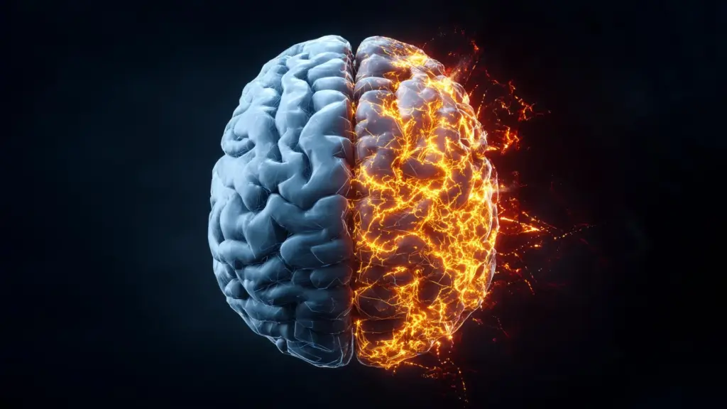
In a significant development for neurology and psychiatry, a team of researchers at the University of Birmingham has identified a promising new strategy to curb harmful brain inflammation. The study, published in the journal Brain, points to the P2X7 receptor as a primary driver of neuroinflammatory processes. By successfully blocking this receptor in human brain tissue, scientists have demonstrated a potential method for mitigating the inflammatory response, a discovery that could lead to the repurposing of existing medications to treat some of the most complex and debilitating brain disorders.
Led by Professor Nicholas Barnes from the University of Birmingham’s College of Medicine and Health, the research team focused on the P2X7 receptor, which acts as a key signaling mechanism in the brain’s immune response. While inflammation is a natural biological process meant to protect the body, persistent or uncontrolled neuroinflammation is a hallmark of numerous neurological and psychiatric conditions. The findings suggest that by targeting this specific receptor, clinicians may eventually be able to dampen the inflammatory damage associated with Alzheimer’s disease, Parkinson’s disease, traumatic brain injury (TBI), depression, and psychosis.
Blocking a Key Driver of Brain Inflammation
The P2X7 receptor has long been a subject of interest in cellular biology due to its role in regulating inflammatory signaling. To better understand how this receptor contributes to the pathology of brain disease, the Birmingham team employed a dual approach, utilizing both live cultures of human brain cells and actual slices of brain tissue harvested during clinical neurosurgical procedures. This methodology allowed the researchers to observe how the receptor influences the release of cytokines—proteins that act as messengers in the body’s inflammatory response.
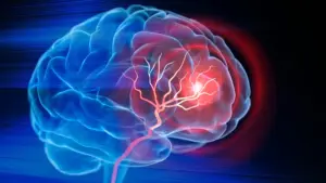
The researchers discovered that P2X7 receptors are essentially "switches" that promote the release of these inflammatory cytokines. When the brain is under stress or injury, these receptors become overactive, leading to an exaggerated inflammatory cascade that can exacerbate tissue damage. By applying a specific antagonist—a drug designed to block the receptor’s activity—the researchers observed a significant decrease in the inflammatory response within the human brain tissue samples.
Professor Barnes emphasized the clinical potential of this discovery, noting that the study provides a roadmap for utilizing existing pharmacological tools in new ways. "This exciting discovery marks a major step toward repurposing existing therapeutics to combat neuroinflammation at its source," Professor Barnes said. "The identification of this receptor could have far-reaching implications for some of the most debilitating and widespread brain disorders such as Alzheimer’s Disease, Parkinson’s and Multiple Sclerosis, or inflammation-linked psychiatric conditions like schizophrenia and depression."
Studying the Brain’s Immune Cells
Central to the research is the role of microglia, the resident immune cells of the central nervous system. Microglia are tasked with monitoring the brain environment, clearing cellular debris, and coordinating the response to injury. However, when these cells become chronically activated, they can contribute to the very damage they are meant to prevent. Studying human microglia has historically been a major obstacle for researchers; once these cells are extracted from the complex environment of the human brain, they typically lose their defining characteristics and functional traits, making it difficult to replicate their behavior in a laboratory setting.
To overcome this hurdle, the University of Birmingham team developed a sophisticated method for converting peripheral blood monocytes—a type of white blood cell—into cells that mirror the behavior of microglia. This cellular transformation is particularly noteworthy because it reflects a process that has been identified as occurring naturally within the human brain during the aging process. By using these monocyte-derived microglia, the researchers created a robust, scalable, and virtually unlimited platform for studying human microglial biology with a level of precision that was previously unattainable.
This innovative model allowed the team to witness how these cells respond to inflammatory signals in real-time. When the researchers induced damage to the microglia-like cells, they were able to use the P2X7 receptor antagonist to interrupt the signaling pathways that would otherwise lead to uncontrolled inflammation.
"Studying human microglia has long been a major challenge," Professor Barnes explained. "Once removed from their native brain environment, they rapidly lose their defining characteristics, likely due to the absence of critical regulatory signals. Our approach involved the use of monocyte-derived microglia, which provide a powerful, scalable, and virtually unlimited platform for studying human microglial biology with unprecedented precision."
From Lab Models to Human Brain Tissue
While the results from the monocyte-derived cells were promising, the ultimate test of the theory’s validity required applying it to actual human brain tissue. By transitioning from their laboratory-grown models to tissue samples obtained during neurosurgical procedures, the researchers were able to confirm that the P2X7 receptor antagonist functioned as predicted in the complex, three-dimensional environment of the human brain.
The successful translation of these results from lab-grown cells to human surgical tissue represents a crucial milestone. It provides the necessary evidence to move toward the next phase of research: clinical trials. This is particularly significant for conditions like traumatic brain injury and certain neurodegenerative diseases where current medical options are severely limited. In many of these cases, the damage to the brain is not just a result of the initial injury or disease progression, but is compounded by the body’s own inflammatory reaction, which can persist long after the initial insult.
The researchers believe that if the P2X7 receptor can be safely targeted in a clinical setting, it could provide a desperately needed therapeutic intervention. The ability to use existing drugs—those that are already known to interact with this receptor—could potentially shorten the time required for development and regulatory approval, offering a faster path to patients in need.
"Having identified the response in the human monocyte-derived microglia, this provided the impetus to translate these findings with human brain obtained following neurosurgical procedures," Professor Barnes stated. "This successful translation means the next stage for this research is the development of clinical trials in patients with neurodegenerative conditions and patients with TBI, where there are no effective pharmacological treatments to reduce the neuroinflammation and arising damage."
As the scientific community looks toward the next steps, the work conducted at the University of Birmingham offers a clear direction for future research. By focusing on the mechanisms of inflammation at a cellular level and leveraging the potential of existing pharmacological agents, the team has opened a new window into the treatment of brain disorders. While clinical trials remain on the horizon, the study establishes a firm foundation for understanding how to manage the inflammatory response in the human brain, potentially transforming the standard of care for millions of patients worldwide. The research serves as a testament to the power of combining innovative cellular modeling with traditional tissue analysis to address some of the most persistent challenges in modern medicine.