






For decades, the scientific community has been preoccupied with the brain’s "hardware"—the intricate web of neurons, the firing of synapses, and the complex expression of genes and proteins. This relentless focus has yielded a remarkable understanding of how our neural circuitry functions. Yet, beneath the surface of these well-studied components lies a critical, often overlooked dimension of neurobiology: the brain’s lipid landscape. These fatty molecules, which serve as the fundamental building blocks for cell membranes, the protective myelin sheaths surrounding nerve fibers, and the signaling pathways essential for cellular communication, have remained largely mysterious, acting as a "dark matter" of brain biology.
A groundbreaking study led by researchers at the Swiss Federal Institute of Technology Lausanne (EPFL) has finally brought this elusive chemical landscape into focus. By creating the first detailed, high-resolution map of lipid distribution in the mouse brain, the team has revealed a highly organized, sophisticated chemical architecture that challenges our traditional understanding of how the brain is structured and how it adapts to life’s major biological transitions.
The Challenge of Mapping the Brain’s Fatty Architecture
The difficulty in studying brain lipids has always been a matter of resolution and technology. While genetic sequencing and protein mapping have advanced rapidly, conventional imaging techniques have struggled to distinguish between the vast array of individual lipid molecules within a tissue sample. Because lipids are so chemically similar to one another, scientists have historically been left with a "blurry picture," unable to pinpoint how lipid composition changes from one anatomical region to another.

"So scientists have had only a blurry picture of how lipids differ from one part of the brain to another," explains Giovanni D’Angelo, professor at The Kristian Gerhard Jebsen Foundation Chair in Nutrition and Metabolism at EPFL. This lack of clarity has been a significant barrier to progress, as it obscured the chemical nuances that likely underpin both healthy brain function and the onset of neurological disorders. D’Angelo emphasizes that this gap is not merely academic; changes in brain lipids have been increasingly linked to a wide spectrum of conditions, ranging from depression to neurodegenerative diseases such as Alzheimer’s.
To address this, a team led by D’Angelo, alongside colleagues Luca Fusar Bassini and Gioele La Manno, set out to construct the "Lipid Brain Atlas." Their work, recently published in the journal Nature, represents a significant shift in how we perceive the brain’s chemical organization.
Constructing the Atlas: 539 Distinct Chemical Zones
The methodology behind the Lipid Brain Atlas is as impressive as its findings. To map the lipids, the researchers utilized mass spectrometry imaging—a sophisticated technique that allows for the identification of specific molecules across thin, precise sections of brain tissue. By employing a laser to scan each slice, location by location, the team was able to capture a high-resolution snapshot of the chemical environment at every point.
Over the course of their study, the researchers examined 172 different types of lipids across 109 brain slices taken from 11 mice. This process generated a massive dataset consisting of approximately 7 million individual measurements. To synthesize this information, the team employed custom-developed machine learning tools, which allowed them to translate raw mass spectrometry data into a comprehensive 3D reconstruction.
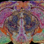
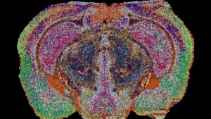
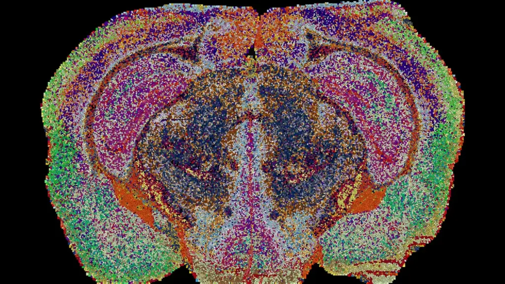
The resulting map identified 539 chemically distinct territories within the brain, which the team has dubbed "lipizones." Each lipizone possesses a unique, identifiable combination of lipids—a chemical signature that is consistent enough to allow researchers to identify the origin of a tissue sample based solely on its lipid profile.
This discovery has profound implications for how we view brain anatomy. While many of these lipizones align perfectly with established anatomical structures or cell types, others revealed entirely new boundaries that were previously invisible to researchers relying on traditional methods. These chemical zones often reveal unexpected relationships between distant clusters of nerve cell bodies and the fibers that connect them.
"In effect, lipids work like postal codes, giving distant but related parts of the brain the same chemical address," D’Angelo notes. This suggests that the chemical environment of the brain is far more interconnected and organized than previously understood, creating a level of functional geography that gene activity alone cannot explain.
Hidden Chemical Patterns and the Complexity of White Matter
Perhaps the most surprising aspect of the Lipid Brain Atlas is its ability to highlight internal variability in regions previously thought to be uniform. A prime example is white matter, the dense tissue composed of myelinated nerve fibers that acts as the brain’s primary communication highway.
For years, white matter was generally treated as a consistent, homogeneous substance. However, the new lipid map indicates otherwise. The atlas reveals that white matter is actually partitioned into numerous distinct lipid zones, suggesting that the glial cells responsible for creating myelin—oligodendrocytes—are far more specialized and chemically diverse than gene expression data would suggest.
"This shows that the cells which make myelin are far more varied in their chemistry than their genes alone would suggest," says Luca Fusar Bassini. This level of chemical diversity implies that the myelin sheath itself might be "tuned" to the specific needs of the underlying nerve fibers, potentially allowing for more nuanced regulation of signal transmission across the brain.
Furthermore, the team identified previously unrecognized chemical boundaries within the choroid plexus, the specialized structure that produces cerebrospinal fluid, as well as in the delicate tissues that line the brain’s internal cavities. These findings suggest that the brain’s chemical organization is a highly dynamic and specialized system, providing a layer of functional control that operates independently of—and perhaps in coordination with—its genetic and anatomical frameworks.
Pregnancy and the Plasticity of Brain Chemistry
Beyond mapping the static structure of the healthy brain, the research team utilized the atlas to explore how the brain’s chemical landscape changes in response to physiological shifts. By examining the brains of pregnant mice, the researchers observed widespread, significant changes in lipid composition across multiple regions.
These changes were found to be more pronounced than the baseline lipid differences typically observed between male and female mice. The cortex, the seat of higher-order cognitive function, exhibited particularly extensive alterations in its lipid profile. Perhaps most tellingly, levels of galactosyl ceramide—a crucial component of myelin—increased sharply across the white matter of the brain.
These results suggest that pregnancy acts as a powerful driver of neurochemical remodeling. The brain appears to be "retuning" its own internal wiring to adapt to the physiological demands of pregnancy, likely optimizing neural connections to meet the body’s changing requirements.
"The Atlas reveals that the brain constantly adjusts its lipids to meet the body’s changing needs," says Gioele La Manno. This discovery highlights the brain’s remarkable plasticity, suggesting that its chemical composition is not a fixed trait but a responsive, adaptive feature that evolves alongside the body’s life stages.
A New Frontier for Disease Research
The Lipid Brain Atlas is now freely available online, inviting the global scientific community to explore the data and integrate it with existing genetic and cellular maps. By providing a detailed, high-resolution reference for healthy mouse brain tissue, the atlas gives researchers a powerful tool for investigating how disruptions in lipid distribution might underpin various pathologies.
While the current map focuses on the mouse brain, the methodology holds great promise for future human studies. As researchers begin to apply these techniques to human tissue, they may be able to identify comparable chemical markers for neurodegenerative diseases like Alzheimer’s or psychiatric conditions like depression.
"What transfers to humans isn’t the map, it’s the method, and the fact that we now know that a healthy brain has a very organized lipid map at all," says Bassini. By establishing that the brain operates according to a highly precise chemical plan, the EPFL team has opened a new chapter in neuroscience—one where the humble lipid is finally recognized as a central player in the complex drama of brain health and function. As the research continues, this atlas will likely serve as a cornerstone for future studies aimed at decoding the chemical language of the mind.