








An unusually detailed, post-mortem examination of an Alzheimer’s patient’s brain has provided scientists with a rare, "side-by-side" look at how clearing harmful protein deposits can fundamentally alter the progression of the disease. The study, which was presented at the 2026 Alzheimer’s Association International Conference and published in JAMA, offers some of the most compelling evidence to date that successfully removing amyloid plaques—the sticky protein clusters that build up outside nerve cells—may actively protect the brain from the secondary, more devastating process of tau protein accumulation.
For decades, the "amyloid hypothesis" has been the cornerstone of Alzheimer’s research, positing that the buildup of beta-amyloid is a primary driver of the neurodegenerative cascade. However, proving that these treatments work in a human brain has been fraught with challenges. This new investigation, conducted by researchers at the Perelman School of Medicine at the University of Pennsylvania, utilized the donated brain tissue of a clinical trial participant to observe, in high resolution, the biological outcomes of long-term anti-amyloid therapy.
A Rare "Goldilocks" Scenario
The study focused on a man who was diagnosed with mild cognitive impairment due to Alzheimer’s in his fifties. He subsequently enrolled in a clinical trial for aducanumab, receiving 30 doses of the medication over a period of four and a half years. Following his death approximately four years after his final dose—and more than a decade after his initial symptoms surfaced—his brain was donated to the Center for Neurodegenerative Disease Research at the University of Pennsylvania’s Department of Pathology and Laboratory Medicine.
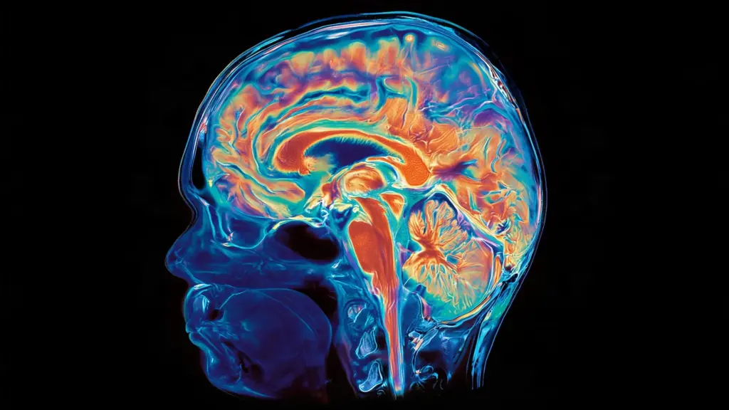
What the researchers discovered upon examination was a remarkable, localized pattern of disease. The brain featured a "patchwork" distribution of pathology that perfectly mirrored imaging scans taken while the patient was still alive. In the gyri—the raised, folded ridges of the brain’s surface—researchers found that the amyloid-clearing treatment had been highly effective. These areas were largely free of amyloid plaques and, crucially, showed very little buildup of tau tangles.
In stark contrast, the sulci—the deep grooves nestled between those ridges—still contained significant amyloid buildup. In these specific areas, the researchers observed high levels of tau tangles, widespread inflammation, and clear evidence of ongoing neurodegeneration.
"Seeing both disease patterns side-by-side in the same brain gave us a rare opportunity to understand how amyloid removal affects other proteins that contribute to Alzheimer’s disease," explained Dr. David Wolk, co-senior author of the study, co-director of the Penn Memory Center, and director of the Penn Alzheimer’s Disease Research Center. "The findings provide some of the clearest human evidence to date that anti-amyloid therapies may limit the accumulation of tau and slow the brain changes that lead to memory loss and cognitive decline."
Dr. Edward B. Lee, the study’s other co-senior author and the John Q. Trojanowski, MD, PhD Professor for Neurodegenerative Disease Research, described the case as a unique "Goldilocks" scenario. Because the treatment had cleared some regions but not others, it created an ideal control environment within a single organ. "Many previous reports have shown either extensive amyloid clearance or limited clearance," Dr. Lee noted. "This allowed us to directly compare what happened next in neighboring brain regions and better understand the relationship between amyloid, tau, and neurodegeneration."
The Relationship Between Amyloid and Tau
To understand the significance of these findings, one must consider the two primary biological culprits in Alzheimer’s disease: beta-amyloid and tau. Beta-amyloid plaques accumulate in the extracellular space between neurons, while tau protein—which normally stabilizes the internal structure of neurons—becomes hyperphosphorylated, detaches from its structural role, and forms toxic "tangles" inside the cells.
For years, the scientific consensus has held that amyloid buildup creates an environment that facilitates the development of tau tangles. Once tau begins to spread, it is widely believed to be the primary driver of the progressive destruction of brain cells, leading to the clinical symptoms of Alzheimer’s: memory loss, confusion, personality changes, and eventually, total cognitive collapse.
While pharmaceutical companies have successfully developed drugs to target amyloid—most notably the now-withdrawn aducanumab, and the currently approved lecanemab (Leqembi) and donanemab (Kisunla)—it has been difficult to definitively prove in human patients that the clearance of amyloid prevents the subsequent tau cascade. Direct evidence has been historically scarce because very few patients who have undergone anti-amyloid treatment have participated in comprehensive post-mortem brain examinations. This study bridges that knowledge gap by demonstrating that in areas where the amyloid was successfully removed, the tau pathology was significantly blunted.
Implications for Future Treatment Strategies
The findings hold profound implications for the current landscape of Alzheimer’s management. If the removal of amyloid can prevent the accumulation of tau, then early intervention becomes the most important factor in long-term cognitive health.
However, researchers are cautious about drawing overly broad conclusions from a single case study. The investigation cannot definitively establish exactly how much amyloid must be removed to achieve permanent protection, nor can it prove that the clearance of plaques was the sole cause of the variance in tau accumulation. Furthermore, the study does not address why some regions of the brain are more resistant to drug delivery than others—a question that remains a significant hurdle in neuropharmacology.
Nevertheless, the study suggests that the benefits of modern Alzheimer’s drugs may be longer-lasting than previously anticipated. Most clinical trials evaluate the efficacy of these medications over a period of 18 months, but the biological impact on tau accumulation might take years to manifest. This research suggests that the protective effects against neurodegeneration may continue to accrue long after the active phase of treatment has concluded.
The Case for Early Intervention
The timing of treatment is perhaps the most critical takeaway from this research. Because beta-amyloid begins to accumulate in the brain years, and sometimes decades, before the first clinical symptoms of Alzheimer’s manifest, there is a growing consensus that the most effective way to combat the disease is to treat it during the preclinical or very early symptomatic stages.
"Patients may accumulate amyloid for years before symptoms appear," said Dr. Christopher A. Brown, an assistant professor of Neurology and the study’s first author. "This case suggests that removing amyloid early may help limit the changes that ultimately damage brain cells. Ongoing trials will help determine whether starting treatment before symptoms begin can provide even greater benefit."
As the scientific community moves forward, the focus is shifting toward identifying the biological mechanisms that allow for more effective amyloid clearance in certain brain tissues and exploring how to replicate those conditions across the entire organ. Researchers are also looking toward a future where patients at high risk for Alzheimer’s could be screened and treated before the "tau cascade" begins, potentially stalling or even preventing the onset of the disease entirely.
The study, which was supported by multiple grants from the National Institutes of Health, highlights the importance of brain donation in the era of targeted Alzheimer’s therapy. By providing a clear window into the structural changes occurring at the cellular level, such research is helping to transform Alzheimer’s from a mysterious, inevitable decline into a condition that, while still formidable, may eventually be managed and even curtailed through precise, early medical intervention.