





Deep within the brainstem lies a tiny, unassuming cluster of nerve cells that has long been considered one of the brain’s most powerful "broadcasting stations." Known as the locus coeruleus (LC)—a name derived from the Latin for "blue place" due to its distinct pigmentation—this region occupies a remarkably small footprint in the human brain. Despite its modest size, it serves as the primary engine for producing norepinephrine (NE), a vital chemical messenger responsible for orchestrating some of the most fundamental aspects of human existence, including attention, learning, heart rate regulation, and the physiological responses to stress and excitement.
For decades, the prevailing consensus among neuroscientists was that the locus coeruleus functioned much like a loudspeaker or a foghorn. The assumption was that the LC broadcast a uniform, monolithic signal across the brain, washing entire neural circuits in norepinephrine to prepare the organism for a "fight or flight" response or to heighten alertness globally. However, a groundbreaking study recently published in the journal Nature has fundamentally challenged this long-held paradigm. Through a sophisticated combination of high-resolution brain imaging, precise neural activity recordings, cutting-edge genetic techniques, and rigorous behavioral experiments in mice, researchers at the Allen Institute have revealed that the locus coeruleus is far more nuanced than previously imagined. Rather than emitting a single, generalized message, the LC utilizes a series of distinct, highly organized pathways to deliver tailored signals to specific destinations, with different subpopulations of neurons reacting uniquely to the experiences of the animal.
A Hidden Brain Network That Sends Precise Messages
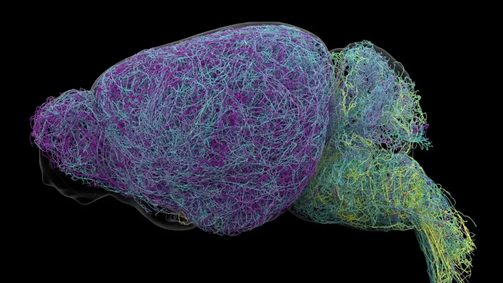
The discovery emerged from a comprehensive effort to map the anatomical connections of individual LC neurons. By tracing these microscopic pathways, the Allen Institute researchers found that the region is not a homogenous blob of cells, but rather a structured communication hub. Different groups of cells within the LC are "hardwired" to communicate with specific, localized regions of the nervous system. This spatial organization ensures that norepinephrine is released only where it is needed, creating a targeted postal network rather than an indiscriminate broadcast.

The researchers identified a clear topographical split in the LC’s architecture. Neurons located in the upper, or dorsal, portion of the structure primarily extend their reach toward the cerebral cortex and the forebrain—areas of the brain involved in complex cognitive processing and executive function. Conversely, cells residing in the lower, or ventral, portion of the LC project their signals primarily to the brainstem and the spinal cord. This structural arrangement provides the brain with a sophisticated mechanism to influence separate and distinct functions simultaneously.
"The findings suggest the brain’s norepinephrine system is far more like a targeted postal network than a foghorn," said Karel Svoboda, director of Neural Dynamics at the Allen Institute and a co-author of the study. This shift in understanding from a "one-size-fits-all" model to a highly compartmentalized system of specialized circuits has profound implications for how we view brain health and pathology.
Why the Discovery Could Matter for Brain Disorders
The implications of this refined map are far-reaching, particularly for the treatment of neurological and mental health conditions. Because norepinephrine is central to the regulation of attention, mood, and physiological arousal, it has long been a primary target for pharmacological interventions. Medications used to manage depression, anxiety, and Attention Deficit Hyperactivity Disorder (ADHD) often aim to modulate norepinephrine levels. Furthermore, the locus coeruleus is historically significant in the study of neurodegenerative disease; it is among the very first regions of the brain to exhibit deterioration in patients with Alzheimer’s disease.
Current therapeutic approaches that influence norepinephrine levels often face the "side effect" hurdle. Because these drugs typically impact multiple brain circuits simultaneously, they can produce beneficial effects in one region—such as improved focus—while causing undesirable physiological responses elsewhere, such as changes in heart rate or blood pressure. By developing a clearer, more granular map of the LC’s specialized pathways, scientists may eventually be able to design next-generation therapies that act with surgical precision. These selective treatments could theoretically target the specific circuits involved in particular symptoms while leaving unrelated neural systems undisturbed. The research, which was supported by the National Institutes of Health’s Brain Research Through Advancing Innovative Neurotechnologies (BRAIN) Initiative, provides the foundational blueprint necessary for this next era of precision neurology.
Scientists Identify Brain Cells Involved in Learning
Beyond the structural anatomy, the researchers uncovered a clear functional division between different groups of LC neurons. In experiments where mice performed decision-making tasks, the researchers observed that dorsal LC neurons—the ones projecting to the cortex—were especially active during the learning process. When the mice were forced to adjust their behavior following an unfavorable result, these neurons showed a distinct increase in activity, suggesting they play a critical role in cognitive flexibility and error correction.
In contrast, the ventral LC neurons exhibited a completely different pattern of behavior. Their activity levels rose shortly before the mice chose to ignore cues that a reward might be available. This suggests that the ventral portion of the LC acts as a gatekeeper for engagement, regulating whether an animal chooses to interact with its environment or remain passive. "What emerged was a clear map: neurons in the dorsal LC that send signals upward to the cortex are involved in learning, whereas neurons in the ventral region projecting downward to the brainstem and spinal cord govern whether animals engage with their environment at all," explained Svoboda. The researchers further confirmed that these functional differences are not merely based on location; they are mirrored by distinct patterns of gene expression, indicating that the neurons in these two regions are biologically specialized for their unique roles.
Researchers Discover a Record-Breaking Neuron
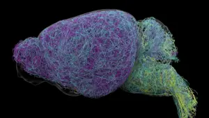
To achieve this level of detail, the study required an unprecedented examination of the mouse brain. The team utilized whole-brain imaging to analyze nearly 35,000 neurons and conducted genetic profiling on approximately 400,000 individual cells. Perhaps most strikingly, the researchers reconstructed the complete physical structure of several exceptionally complex nerve cells, revealing that LC neurons possess remarkably long axons—the slender, thread-like projections that carry electrical impulses to other parts of the nervous system.
The average length of these axons was measured at approximately 35 centimeters. However, one specific axon reached a staggering 70.32 centimeters, marking it as one of the longest single neurons ever documented in a mouse. "This neuron, like many others that we studied, supplies NE to a very large volume of the cerebral cortex. For the brain, this is highly unusual. Most neurons are more specific in their targets," said Jeremiah Cohen, a scientist at the Allen Institute and a co-author of the study. Despite this immense length and reach, the neuron does not function as a blanket distributor. It remains highly selective, ignoring the cerebellum, brainstem, and spinal cord. This reinforces the central theme of the study: even at the scale of a record-breaking, long-range connection, the brain maintains a high level of organizational control over its chemical signals.
A New Understanding of How the Brain Learns
Finally, the study identified fascinating parallels between the norepinephrine system and the brain’s dopamine signaling system. Dopamine is a well-known messenger that facilitates communication within the basal ganglia, a region essential for habit formation and reward processing. Scientists have long hypothesized that norepinephrine and dopamine work in tandem to support various forms of learning. The new evidence regarding the locus coeruleus suggests that these systems may interact to allow the brain to acquire and integrate complex, abstract concepts simultaneously.
By revealing the intricate, specialized, and physically vast connections of the locus coeruleus, this research provides a far more detailed picture of how the mammalian brain coordinates learning and environmental engagement. While these findings were generated using mouse models, they serve as a critical bridge toward understanding the human brain. By shifting the perspective from a generalized broadcast system to a precise, circuit-based network, this work offers a roadmap for developing more effective, targeted treatments for the millions of people living with neurological and psychiatric conditions. As research continues, the "blue place" in the brainstem promises to yield even more insights into the complex mechanics of human cognition.