








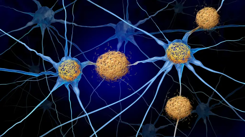
Protecting brain cells from destruction remains one of the most formidable hurdles in the global effort to treat Alzheimer’s disease and a spectrum of related neurodegenerative conditions collectively known as tauopathies. These disorders are characterized by the accumulation of abnormal tangles of a protein called tau within the brain’s architecture. As these tangles proliferate, nearby neurons—the fundamental building blocks of thought and memory—inevitably begin to succumb and die. For decades, the prevailing scientific consensus focused on the tau tangles themselves as the primary architects of this destruction. However, a growing body of evidence suggests a more nuanced and potentially hopeful reality: the tau protein may not be the sole culprit behind the loss of brain cells, memory, and cognitive independence. Instead, the immune system’s misdirected reaction to these protein accumulations appears to play a significantly more destructive role than previously understood.
A breakthrough study conducted by researchers at the Washington University School of Medicine in St. Louis has identified a promising strategy for interrupting this harmful immune response. By targeting the mechanism that allows immune cells to infiltrate the brain, scientists have successfully preserved brain tissue and cognitive function in animal models, even while the underlying protein tangles remained present. The findings, which were published on September 28 in the journal Neuron, offer a potential shift in how clinicians might one day approach the treatment of devastating neurodegenerative diseases.
The Mechanism of Immune Infiltration
The research team, led by senior author David M. Holtzman, MD, the Barbara Burton and Reuben M. Morriss III Distinguished Professor in the Department of Neurology at WashU Medicine, sought to understand how the immune system—designed to protect the body—ends up facilitating the destruction of brain cells. In studies involving mice engineered to exhibit tau accumulation similar to that seen in human Alzheimer’s patients, the researchers focused on a protein called CXCR3. This protein acts as a gatekeeper, helping immune cells known as T cells migrate from the bloodstream into the brain.
In healthy conditions, the immune system is highly regulated. However, in the context of tauopathies, the environment of the brain becomes inflammatory. The researchers discovered that by using an antibody to block CXCR3, they could effectively disrupt the pathway that guides T cells into the brain’s sensitive regions. Following a treatment regimen that lasted several months, the number of T cells found within the brains of the treated mice had fallen by approximately 50% compared to the control group.
The results were striking in their clinical significance. The mice that received the antibody therapy retained roughly 40% more tissue in key brain regions responsible for memory and executive function compared to their untreated counterparts. Furthermore, the treated animals demonstrated superior performance on standardized memory tests. Perhaps most remarkably, these improvements in tissue preservation and cognitive function were achieved despite the fact that the total amount of accumulated tau in the brain remained unchanged. This discovery fundamentally challenges the idea that reducing protein tangles is the only path to preventing neurodegeneration.
"In tauopathies, including Alzheimer’s disease, there’s no treatment right now that actually decreases neurodegeneration," Dr. Holtzman observed. "If we can show that we’re really decreasing brain cell death, it’s certainly worth trying to pursue that pathway to prevent the most devastating consequences of these diseases."
Rethinking the Role of the Immune System
To understand the significance of this finding, it is necessary to examine the current landscape of Alzheimer’s treatment. Modern therapeutic approaches have primarily focused on amyloid, a different protein that forms plaques between neurons during the early stages of the disease. Medications such as lecanemab and donanemab, which have gained regulatory approval, are designed to clear these amyloid deposits. While these drugs can effectively slow cognitive decline, they have not been proven to prevent the actual death of brain cells. Additionally, these amyloid-targeting therapies offer no relief for primary tauopathies—conditions where tau builds up without the presence of amyloid plaques. Alzheimer’s is classified as a secondary tauopathy because both amyloid and tau are present, but the mechanisms of damage may differ significantly.
Dr. Holtzman’s laboratory has spent years investigating whether the immune system might hold the key to addressing the gaps left by current therapies. In a 2023 study published in the journal Nature, his team discovered that T cells were not merely bystanders; they were actively migrating into the brains of mice with abnormal tau, concentrating specifically in the regions where tau tangles were most dense. Once there, these T cells contributed significantly to the neurodegenerative process. A follow-up study published earlier this month in Nature Neuroscience added another critical piece to the puzzle, revealing that the signals directing these T cells originate in the lymph nodes located outside the brain, rather than within the brain itself.
Identifying the Chemical Roadmap
Despite these breakthroughs, the researchers still faced the challenge of understanding exactly how activated T cells navigated the complex biological terrain to reach the brain. Immune cells typically navigate the body by following chemical signals known as chemokines. Earlier experiments in the Holtzman lab had identified that mice with tau accumulation exhibited unusually high levels of a chemokine called CXCL10. Significantly, other researchers had reported similar elevations of CXCL10 in human patients suffering from Alzheimer’s disease.
Activated T cells carry a surface receptor known as CXCR3, which functions like a biological antenna, allowing the cells to lock onto and follow the signals emitted by CXCL10. The researchers hypothesized that this CXCL10-CXCR3 pathway was essentially a chemical "roadmap" guiding harmful T cells into the brain. To test this, the team, which included co-senior author Jason Ulrich, PhD, a professor of neurology at WashU Medicine, and first author Joshua T. Emmerson, PhD, a postdoctoral researcher, studied mice that developed tau deposits mimicking those in human Alzheimer’s patients.
The experiments were conclusive: when the mice lacked either the CXCL10 signal or the CXCR3 receptor on their T cells, the cells could not infiltrate the brain. This protective effect remained stable even when the researchers deliberately introduced inflammation to stimulate an immune response. With the pathway identified, the team shifted their focus to whether blocking this pathway with an antibody could prevent the actual onset of brain damage.
Preserving Memory and Brain Integrity
In the experimental treatment phase, the researchers utilized young mice that had begun to develop tau deposits but had not yet reached the stage of significant neurodegeneration. For a period of three and a half months, the mice were treated with the CXCR3-blocking antibody every five days.
The outcome was a clear validation of the hypothesis. By the end of the study, the treated mice exhibited half the number of infiltrating T cells in their brains compared to the untreated group. More importantly, there was a visible reduction in neuronal injury and a preservation of approximately 40% more brain tissue in regions linked to memory. The behavioral tests confirmed that this tissue preservation translated into improved cognitive function.
A critical takeaway from this study, with major implications for future drug development, is that the antibody did not need to enter the brain to be effective. The antibody reached the boundary of the brain but remained outside the blood-brain barrier. This is a "big deal" in the field of neurology, according to Dr. Holtzman, because the blood-brain barrier is notoriously difficult to cross, and many promising drug candidates fail in clinical trials simply because they cannot reach their target tissue in sufficient concentrations.
"For this therapeutic approach, if it is safe, you wouldn’t have to design the drug to get into the brain—which is a big deal since most molecules don’t cross the blood-brain barrier well—and you don’t have to get rid of the tau to get this therapeutic effect," Dr. Holtzman explained.
A New Horizon for Alzheimer’s Therapy
While the results in animal models are highly encouraging, the researchers emphasize that further study is required to determine the safety and efficacy of this approach in human patients. However, the scientific community may already have a head start. Dr. Holtzman suggested that existing medications used to regulate T cells in other diseases—such as multiple sclerosis and other autoimmune conditions—could serve as a foundation for clinical investigations.
This strategy represents a potential paradigm shift in the treatment of neurodegeneration. By moving beyond the exclusive focus on protein deposits and instead targeting the immune system’s damaging secondary responses, researchers may be able to offer a more effective way to protect neurons and preserve memory. As Dr. Holtzman noted, tauopathies are not traditionally categorized as autoimmune disorders, yet this study demonstrates for the first time in an animal model that they can respond to specific T-cell-modulating therapies.
The research received broad support from organizations including the BrightFocus Foundation, the National Institutes of Health, the GHR Foundation, and the Carol and Gene Ludwig Initiative in Neuroimmunology Research, among others. As the field moves forward, the possibility of protecting the brain from the collateral damage of the immune system remains a compelling, hopeful, and potentially transformative frontier in the fight against Alzheimer’s disease and related tauopathies.