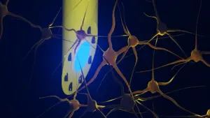










In a groundbreaking study that challenges decades of conventional wisdom, scientists have uncovered a surprising and potentially reversible cause for the chronic sleep disturbances that plague millions of individuals living with Alzheimer’s disease. By shifting the focus away from the amyloid plaques that have long dominated Alzheimer’s research, investigators at the University of Kentucky have identified the brain’s own immune system—specifically cells known as microglia—as a primary driver of sleep loss.
The research, published in the journal Alzheimer’s & Dementia, reveals that when these immune cells overreact to the presence of amyloid plaques, they initiate an inflammatory cascade that keeps the brain in a state of hyper-arousal. Most significantly, the team demonstrated that by temporarily suppressing these cells, they could restore over two hours of sleep per day in mouse models, even while the amyloid plaques remained untouched in the brain. This "paradigm-shifting" discovery suggests that it may be possible to treat one of the most debilitating symptoms of the disease without needing to clear the plaques themselves, offering a new frontier in clinical intervention.
How the Brain’s Immune System Disrupts Sleep
To understand the mechanics of this discovery, it is helpful to visualize the brain’s response to injury. Under normal circumstances, microglia act as the brain’s "housekeepers." They are specialized immune cells tasked with clearing cellular debris, monitoring for injury, and maintaining an environment conducive to healthy neuronal function. However, in the presence of Alzheimer’s pathology—specifically the accumulation of sticky amyloid protein fragments known as plaques—this defensive response can become dangerously dysregulated.

Shannon L. Macauley, Ph.D., an associate professor of physiology at the University of Kentucky College of Medicine and lead investigator of the study, likens the phenomenon to a domestic emergency gone wrong. "Imagine a small fire breaking out in a kitchen," Macauley explains. "Instead of containing the flames, the building’s sprinkler system activates everywhere, flooding the entire house and causing widespread damage."
In the Alzheimer’s brain, microglia function similarly to that overactive sprinkler system. When they detect amyloid plaques, they do not simply neutralize the threat. Instead, they release a deluge of inflammatory signaling molecules that interfere with the intricate neural circuits responsible for healthy sleep. "Basically, we showed that it is not the plaques themselves, or solely dysfunctional neurons, that cause sleep loss but actually microglia," Macauley notes. "Microglia are immune cells that, when they respond to plaques, kick off this elaborate cascade of inflammation, as if the microglia are partying all night, and keeping the brain awake."
Tracking Alzheimer’s Sleep Changes Inside the Brain
The research team, which included first author Nicholas J. Constantino, Ph.D., a recent doctoral graduate, employed a sophisticated methodology to differentiate between normal aging and Alzheimer’s-related pathology. They compared two cohorts of mice: one group genetically predisposed to develop amyloid plaques and a control group of "wild-type" mice that underwent the standard, healthy aging process.
The researchers monitored these subjects at two critical life stages: six months, when plaques first begin to emerge, and 18 months, when the disease model manifests advanced neurodegeneration. To capture the nuance of sleep architecture, the team fitted the mice with advanced recording devices that utilized electroencephalography (EEG) and electromyography (EMG). While the EEG provided data on the rhythmic electrical activity of the brain—allowing researchers to distinguish between wakefulness, dreaming, and deep, restorative sleep—the EMG recorded muscle activity, providing a complete picture of the animals’ sleep-wake cycles.
To visualize the physical relationship between the plaques and the immune response, the team utilized light-sheet microscopy. By rendering biological tissue transparent and scanning it with a precise, thin laser, they were able to create high-resolution, three-dimensional maps of the brain. This allowed them to pinpoint exactly where microglia were clustering around plaques and where the most intense inflammatory responses were occurring.
The Impact of Immune Cell Depletion
The pivotal moment in the research arrived when the team sought to test the causal link between microglial inflammation and sleep loss. They administered Pexidartinib (PLX3397), a medication originally developed for oncology that specifically targets the survival signals required by microglia. By including this in the mice’s food for a two-week period, the team was able to temporarily eliminate approximately 87% of the brain’s microglia.
The results were immediate and striking. The mice, which had been suffering from significant sleep disruption, gained more than two hours of sleep per day. Crucially, this restored sleep was not merely "light" sleep; the mice experienced more time in non-rapid eye movement (NREM) sleep—the deep, restorative phase essential for physical repair, memory consolidation, and the clearing of metabolic waste products from the brain.
To analyze the underlying brain activity during this transition, the team used a method called Fitting Oscillations and One Over Frequency (FOOOF). This mathematical approach allowed them to separate rhythmic brain waves from "background noise," essentially revealing whether the brain’s internal engine was still "idling" at high speed when it should have been resting. The reduction in microglial activity essentially "calmed" the brain, allowing it to transition into a natural, restful state.
Challenging the "Plaque-First" Model
Perhaps the most surprising finding of the study was the "ceiling effect" observed as the disease progressed. The researchers had long assumed that as the burden of amyloid plaques increased, sleep disruption would worsen in a linear fashion. However, the data told a different story. The mice developed significant sleep problems early in the disease process, but those problems did not escalate as the plaque count doubled or tripled over the subsequent months.
"I expected that as plaque burden became more severe, sleep disruption would also worsen," said Constantino. "The disruptions in sleep and cortical EEG activity that occur at 6 months, when plaques first emerge, did not worsen by 18 months, despite more than double the amount of plaque burden." This suggests that the inflammatory response is triggered early and reaches a peak intensity, after which additional plaque accumulation does not necessarily lead to further sleep loss. This revelation provides a vital clue as to why sleep disturbances are often one of the earliest clinical indicators of Alzheimer’s, potentially appearing years before significant memory decline occurs.
Furthermore, the study highlighted that Alzheimer’s does not affect all sleep stages equally. While normal aging typically results in a reduction of REM sleep, Alzheimer’s pathology selectively targets NREM sleep. Given the critical role of NREM sleep in "washing out" toxins from the brain, this suggests a dangerous, self-reinforcing cycle: the disease disrupts deep sleep, and the lack of deep sleep prevents the brain from clearing the very waste products that drive the pathology.
Toward Clinical Translation and Future Research
While the results in mice are highly encouraging, the researchers are careful to emphasize that they do not yet have a direct clinical application for humans. Removing 87% of a person’s microglia is not a viable long-term treatment strategy, as these cells are essential for maintaining a healthy brain environment. The current goal for the team at the University of Kentucky is to find ways to modulate or "calm" these cells rather than eliminating them entirely.
"We are currently looking at whether we can tweak microglial activity to prevent that ‘all-night party’ of inflammation," says Macauley. Her lab is investigating existing, FDA-approved medications, such as the diabetes drug Metformin and certain anti-seizure medications like Stiripentol, for their potential to alter how microglia use energy. By shifting the metabolic profile of these cells, the team hopes to prevent the inflammatory cascade without compromising the cells’ protective roles.
Additionally, the study offers a secondary benefit: the potential for more accessible diagnostics. Because the team identified specific electrical signatures in the EEG data associated with Alzheimer’s pathology, they envision a future where portable, affordable EEG devices could be used to screen for these biomarkers in community settings. Such a tool could be transformative, particularly in rural or underserved areas where access to expensive, invasive neuroimaging is limited.
As the research progresses, the team continues to embody the "calculated risk-taking" culture that Macauley cultivates in her lab. By questioning the established narrative and following the data, the group has successfully reframed the Alzheimer’s sleep problem from a hopeless consequence of plaque accumulation to a potentially manageable inflammatory condition. While much work remains to translate these findings to human patients, the discovery provides a beacon of hope for improving the quality of life, cognitive health, and daily function of those living with the early stages of Alzheimer’s disease.
The research was supported by the National Institute on Aging and the National Institute of General Medical Sciences of the National Institutes of Health, as well as the Cure Alzheimer’s Fund and the Coins for Alzheimer’s Research Trust (CART). As the team moves forward, their focus remains on the ultimate goal: developing therapies that can safely intervene in the inflammatory process to restore the restorative sleep that is so vital to brain health.