







Deep within the brainstem lies a tiny, unassuming cluster of nerve cells that wields an influence far greater than its physical size would suggest. Known as the locus coeruleus (LC)—a name derived from the Latin for "blue place" due to its distinct appearance in unstained brain tissue—this region has long been recognized as the brain’s primary manufacturer of norepinephrine (NE). This vital chemical messenger acts as a physiological conductor, orchestrating the body’s heart rate, stress responses, attention, and the fundamental mechanics of learning. For decades, the prevailing consensus among neuroscientists was that the LC functioned much like a broadcast loudspeaker or a foghorn, releasing a uniform wave of norepinephrine to saturate large swaths of the brain simultaneously.
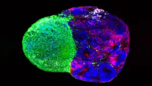
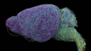
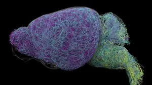
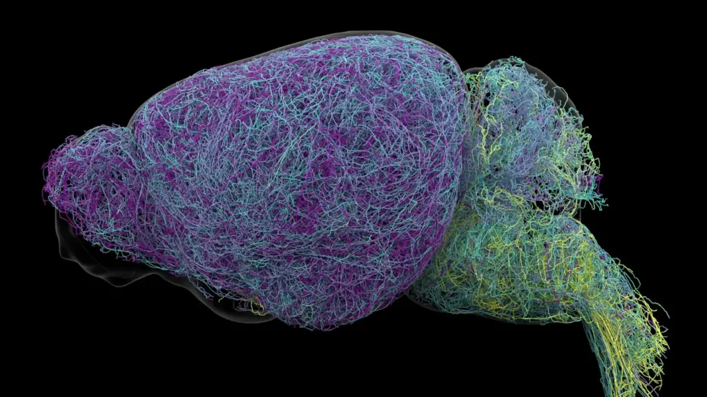
However, a groundbreaking study published in the journal Nature has fundamentally upended this long-standing assumption. Researchers at the Allen Institute, utilizing an advanced suite of brain imaging, high-resolution neural recordings, and sophisticated genetic mapping, have discovered that the LC is not a monolithic broadcaster. Instead, it functions as a highly precise, targeted postal network. By tracing the anatomical and functional architecture of the LC in mice, the research team revealed that this region communicates through several distinct, specialized pathways, delivering nuanced signals to specific destinations rather than a single, generalized message.
A Hidden Brain Network That Sends Precise Messages
The discovery suggests that the brain’s norepinephrine system is far more organized than previously imagined. Through rigorous mapping of LC connections, researchers identified that different subpopulations of neurons are hardwired to communicate with specific regions of the nervous system. This structural arrangement allows the brain to exert granular control over various physiological and cognitive functions, effectively debunking the idea that the locus coeruleus operates as a diffuse, indiscriminate signal generator.

The internal architecture of the LC appears to be organized by geography. Neurons located in the upper, or dorsal, portion of the region primarily extend their reach toward the cerebral cortex and the forebrain, areas associated with complex thought and decision-making. In contrast, cells situated in the lower, or ventral, portion of the LC predominantly project their signals downward to the brainstem and the spinal cord. By segregating these pathways, the brain ensures that different components of the LC can influence separate, independent functions, providing a level of regulatory sophistication that a "foghorn" model could never achieve.
"The findings suggest the brain’s norepinephrine system is far more like a targeted postal network than a foghorn," noted Karel Svoboda, director of Neural Dynamics at the Allen Institute and a co-author of the study. This shift in understanding from a broadcast system to a directed network provides a new framework for viewing how the brain manages state-dependent behavior and environmental engagement.
Why the Discovery Could Matter for Brain Disorders
The implications of this research extend far beyond basic neurobiology; they reach into the heart of clinical medicine and the treatment of neurological and mental health conditions. Because norepinephrine is a central regulator of mood and attention, it has long been a primary target for pharmacological interventions. Medications for depression, anxiety, and Attention Deficit Hyperactivity Disorder (ADHD) often attempt to modulate norepinephrine levels to improve patient outcomes.
However, current pharmaceutical approaches are frequently broad-spectrum, affecting multiple brain circuits simultaneously. This lack of specificity often results in a "double-edged sword" effect: a drug might successfully alleviate one set of symptoms in a specific region of the brain while inadvertently triggering unwanted side effects in another. With this newfound map of the LC’s specialized pathways, scientists have a better blueprint for developing next-generation therapies. By targeting specific neural circuits rather than the entire norepinephrine system, researchers hope to design treatments that are more precise, effective, and free from the systemic side effects that plague current medications.
The relevance of this work is further underscored by the progression of neurodegenerative diseases. Neurons in the locus coeruleus are among the first to show signs of deterioration in Alzheimer’s disease. Understanding the precise roles and connectivity of these cells may offer new insights into why this region is so vulnerable and how its early decline contributes to the cognitive and behavioral symptoms observed in patients. This research, supported by the National Institutes of Health’s Brain Research Through Advancing Innovative Neurotechnologies (BRAIN) Initiative, provides a foundation for more nuanced therapeutic strategies.
Scientists Identify Brain Cells Involved in Learning
Beyond the structural mapping, the researchers uncovered functional distinctions that clarify how the LC contributes to cognitive flexibility. The team found that the dorsal LC neurons, which project to the cortex, play a critical role in the learning process. When mice were tasked with making decisions and subsequently had to adjust their choices following unfavorable results, these specific neurons demonstrated a marked increase in activity. This suggests that the dorsal LC is intimately involved in the feedback loops required for learning and error correction.
In contrast, neurons in the ventral LC exhibited a completely different behavioral signature. Their activity spiked shortly before the mice chose to ignore environmental cues that signaled the potential for a reward. This pattern of activity suggests that the ventral LC acts as a gatekeeper, regulating whether an animal decides to engage with its surroundings or ignore incoming information.
"What emerged was a clear map: neurons in the dorsal LC that send signals upward to the cortex are involved in learning, whereas neurons in the ventral region projecting downward to the brainstem and spinal cord govern whether animals engage with their environment at all," explained Svoboda. "We also showed that these anatomical differences are mirrored by distinct gene expression patterns." This indicates that the distinction between these two groups of neurons is not merely physical; they are genetically programmed to perform different tasks, further reinforcing the idea of a specialized, compartmentalized system.
Researchers Discover a Record-Breaking Neuron
The scale of the study was immense, involving the examination of nearly 35,000 neurons through whole-brain imaging and the genetic profiling of almost 400,000 individual cells. During this meticulous reconstruction process, the team stumbled upon a startling structural finding: the presence of exceptionally long axons. While axons are the cables that allow nerve cells to transmit information, the lengths observed in the LC were extraordinary.
On average, LC axons measured approximately 35 centimeters long. Even more remarkable was the discovery of a single neuron with a reconstructed length of 70.32 centimeters—a measurement that establishes it as one of the longest single neurons ever documented in a mouse. Despite its massive reach, the neuron remained highly selective in its targets, supplying norepinephrine to specific volumes of the cerebral cortex while pointedly ignoring other regions like the cerebellum and brainstem.
"This neuron, like many others that we studied, supplies NE to a very large volume of the cerebral cortex. For the brain, this is highly unusual. Most neurons are more specific in their targets," said Jeremiah Cohen, an Allen Institute scientist and study co-author. The existence of these sprawling yet selective structures emphasizes that even when a neuron covers a large area, its influence is dictated by precise, curated connections.
A New Understanding of How the Brain Learns
The researchers also drew parallels between the norepinephrine system and the brain’s dopamine system, which is well-known for its role in the basal ganglia and habit formation. There is growing evidence that dopamine and norepinephrine may act in concert to support learning, potentially allowing the brain to process multiple, abstract concepts simultaneously.
By unraveling the intricate connections, specialized functions, and the surprising physical architecture of the locus coeruleus, this study offers a significantly more detailed picture of how the brain manages information. While the research was conducted in mice, the fundamental principles of neural circuit organization identified here are likely to inform our understanding of the human brain. As scientists continue to map the complex geography of the mind, the discovery that the "blue place" is actually a hub of highly organized, specialized communication marks a significant leap forward in the quest to treat brain disorders at their source.