





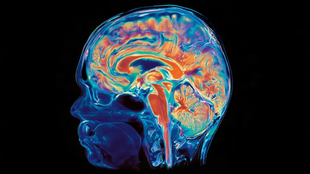
For generations, the scientific community has operated under a foundational assumption: the brain is a single, unified organ that shares a common developmental origin. Whether considering the complex architecture of the human forebrain or the rhythmic, life-sustaining mechanisms of the hindbrain, the prevailing model suggested that all these regions traced their lineage back to a single progenitor cell population during the earliest stages of embryonic growth.
However, new research led by Stanford Medicine is challenging this long-held dogma. According to a study published on September 18 in Nature Neuroscience, the human brain is not a singular developmental entity. Instead, it appears to be a composite structure built from two entirely distinct nervous systems that evolved separately over hundreds of millions of years before eventually converging to form the organ we recognize today. This discovery not only rewrites our understanding of neurobiology but also offers a potential breakthrough for scientists struggling to model and treat devastating neurological disorders like ALS and spinal muscular atrophy.
Two Evolutionary Paths to One Brain
The adult human brain is conventionally divided into three primary regions: the forebrain, the midbrain, and the hindbrain. The forebrain is the seat of our most complex cognitive faculties—the regions responsible for language, abstract reasoning, consciousness, and the capacity for self-reflection. In contrast, the hindbrain, often referred to as the brain stem, is the silent engine of survival. It governs the automatic, involuntary processes that keep us alive, such as regulating the heartbeat, managing sleep cycles, controlling respiration, and processing hunger. Furthermore, the hindbrain houses the neural circuits that control the muscles of the face, tongue, and throat, which are essential for fundamental survival tasks like swallowing and speaking.
Despite the hindbrain’s critical importance, researchers have faced a persistent and frustrating hurdle: the inability to successfully grow hindbrain neurons in a laboratory setting. For decades, attempts to replicate these cells using stem cell technology have consistently failed, leaving scientists without effective models to study diseases that target the brain stem.
Kyle Loh, PhD, an associate professor of developmental biology at Stanford Medicine and the senior author of the new study, notes that this limitation has been a significant barrier in medical research. "We’ve shown for the first time that the front of the brain arises from a totally different progenitor cell than the back of the brain," Loh said. "Our discovery means that we can now grow neurons from the back of the brain, the hindbrain, in a petri dish and study their functions."
The research team, which included co-first authors and graduate students Carolyn Dundes and Rayyan Jokhai, posits that previous failures to culture hindbrain neurons were likely due to a fundamental misunderstanding of embryonic development. Scientists were attempting to "coax" progenitor cells destined for the forebrain or midbrain to become hindbrain cells—a biological impossibility, as the two populations follow distinct genetic blueprints from the very first moments of life.
The Blueprint of Early Development
The key to this discovery lay in examining the earliest stages of gastrulation, the phase in embryonic development where the basic body plan is established. Jokhai and Dundes discovered that the hindbrain does not emerge as a secondary branch of a single developmental pathway. Rather, it follows a parallel, independent trajectory from the very beginning.
To map this process, the team studied developing mouse embryos, where they identified two distinct populations of neural progenitor cells. The first population expresses a gene known as Otx2 and is programmed specifically to form the forebrain and midbrain. A second, entirely separate population expresses the gene Gbx2 and is destined to form the hindbrain. The researchers observed that these two populations never overlap, maintaining their distinct identities even at the earliest stages of development.
This separation is reinforced at the level of chromatin—the complex material that packages DNA and dictates which genes remain accessible or inaccessible to the cell. The anterior neural ectoderm (the precursor to the forebrain and midbrain) and the posterior neural ectoderm (the precursor to the hindbrain) possess fundamentally different chromatin configurations. This chemical signature effectively locks the cells into their respective developmental fates from the start.
"In stem cell biology, people are always fixated with creating the end cell type, like the neuron," Jokhai explained. "But it’s important to begin at the earliest stages of embryonic development. Our careful attention to that early time point allowed us to find this fundamental split in brain development."
A Deep Evolutionary History
The implications of this dual-origin brain extend far beyond the laboratory. By examining the evolutionary record across 550 million years, the researchers found that this two-origin arrangement is not a recent adaptation, but an ancient legacy. The study identified the same dual-pathway pattern in organisms as diverse as chickens, zebrafish, and acorn worms—a marine invertebrate that shares a distant common ancestor with humans.
The team even looked as far back as jellyfish, which diverged from the human lineage roughly 600 to 700 million years ago. These organisms possess two separate nervous systems located at opposite ends of their bodies, suggesting that the modern vertebrate brain may have emerged when evolution physically forced two separate, primordial neural systems into close proximity.
"Our research suggests that evolution took two existing neural systems and pushed them together spatially," Loh said. "Having the brain as one organ would probably be more efficient, but we rely on this primordial way to make the brain as two separate pieces."
Jokhai echoed this sentiment, noting the counterintuitive nature of the findings. "I was surprised at our findings because the word ‘brain’ implies a contiguous organ that likely has a singular origin. But even 500 million years ago, there were these separate neural systems, which now almost operate as one, which is very cool."
New Frontiers for Neurological Medicine
The ability to finally isolate and grow human hindbrain motor neurons using pluripotent stem cells marks a transformative moment for clinical research. For the first time, scientists have a reliable, scalable model to investigate the mechanisms of diseases that specifically target the brain stem, most notably amyotrophic lateral sclerosis (ALS) and spinal muscular atrophy (SMA).
SMA is a devastating genetic disorder and a leading cause of death in children under the age of one. ALS, which typically strikes in mid-to-late adulthood, causes the progressive degradation of both forebrain and hindbrain function. In both conditions, specific hindbrain neurons begin to fail, eventually stripping patients of the ability to swallow or breathe. Because these neurons were previously impossible to cultivate in the lab, research into their failure was largely restricted to animal models or indirect study. Now, researchers can observe these processes directly in human cells.
Furthermore, this discovery may have implications for metabolic research. The hindbrain houses the neural circuits responsible for regulating hunger—systems that are currently the primary targets for emerging weight-loss drugs like semaglutide. With a better understanding of how these circuits form, researchers may be able to refine treatments for obesity and other metabolic conditions.
As the team looks toward the future, they aim to apply these same techniques to investigate the developmental origins of the spinal cord and to untangle the precise ways in which neurodegenerative diseases disrupt the hindbrain. By moving away from the "single-brain" model and embracing the reality of a dual-origin system, scientists are opening a new frontier in regenerative medicine.
"Now we have a model to better understand these devastating diseases, and work toward regenerative therapies for them," Jokhai said. "This is a very exciting new frontier in brain research."
The study, which included contributions from researchers at the California Institute of Technology and the University of California, San Francisco, was supported by a wide array of institutions, including the National Institutes of Health, the National Science Foundation, and the California Institute for Regenerative Medicine, among others.